. 2015 Sep 1;309(5):F456-63.
doi: 10.1152/ajprenal.00631.2014. Epub 2015 Jul 1.

Calmodulin and CaMKII modulate ENaC activity by regulating the association of MARCKS and the cytoskeleton with the apical membrane

Affiliations

Calmodulin and CaMKII modulate ENaC activity by regulating the association of MARCKS and the cytoskeleton with the apical membrane

Abdel A Alli et al. Am J Physiol Renal Physiol. .

Abstract

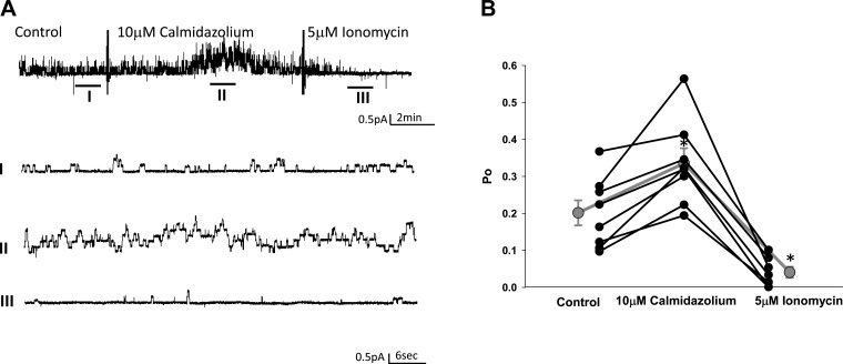
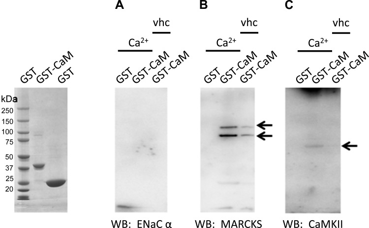
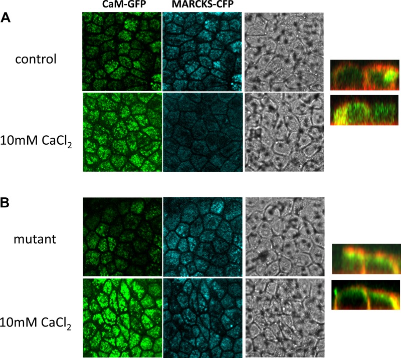
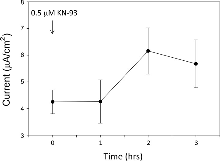
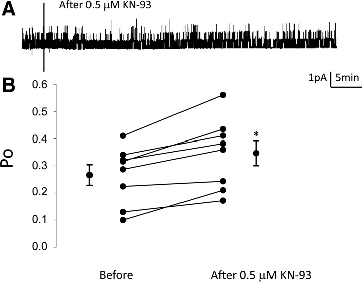
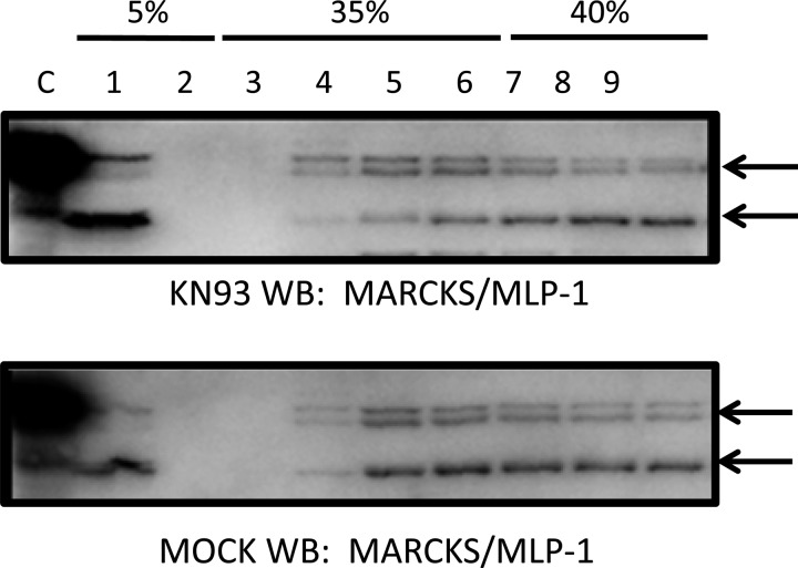
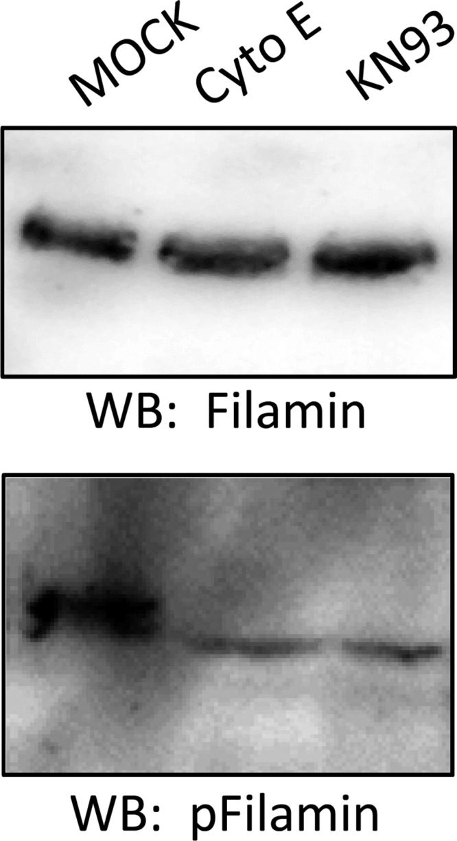
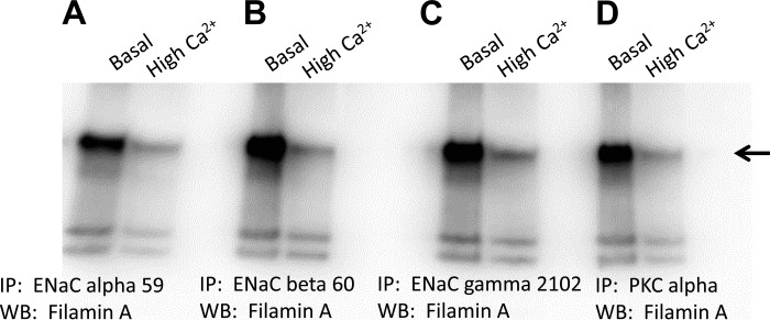
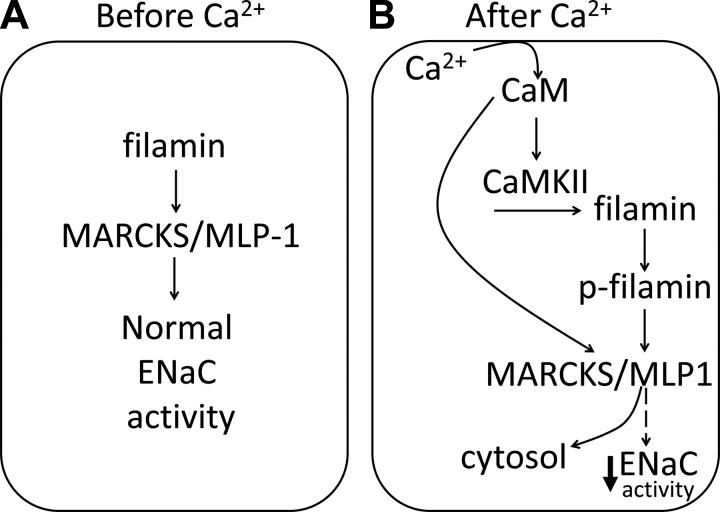
Phosphatidylinositol bisphosphate (PIP2) regulates epithelial sodium channel (ENaC) open probability. In turn, myristoylated alanine-rich C kinase substrate (MARCKS) protein or MARCKS-like protein 1 (MLP-1) at the plasma membrane regulates the delivery of PIP2 to ENaC. MARCKS and MLP-1 are regulated by changes in cytosolic calcium; increasing calcium promotes dissociation of MARCKS from the membrane, but the calcium-regulatory mechanisms are unclear. However, it is known that increased intracellular calcium can activate calmodulin and we show that inhibition of calmodulin with calmidazolium increases ENaC activity presumably by regulating MARCKS and MLP-1. Activated calmodulin can regulate MARCKS and MLP-1 in two ways. Calmodulin can bind to the effector domain of MARCKS or MLP-1, inactivating both proteins by causing their dissociation from the membrane. Mutations in MARCKS that prevent calmodulin association prevent dissociation of MARCKS from the membrane. Calmodulin also activates CaM kinase II (CaMKII). An inhibitor of CaMKII (KN93) increases ENaC activity, MARCKS association with ENaC, and promotes MARCKS movement to a membrane fraction. CaMKII phosphorylates filamin. Filamin is an essential component of the cytoskeleton and promotes association of ENaC, MARCKS, and MLP-1. Disruption of the cytoskeleton with cytochalasin E reduces ENaC activity. CaMKII phosphorylation of filamin disrupts the cytoskeleton and the association of MARCKS, MLP-1, and ENaC, thereby reducing ENaC open probability. Taken together, these findings suggest calmodulin and CaMKII modulate ENaC activity by destabilizing the association between the actin cytoskeleton, ENaC, and MARCKS, or MLP-1 at the apical membrane.

Keywords: CaMKII; ENaC; MARCKS; calcium; filamin.


Figures

Fig. 1.
Fig. 1.
Single-channel patch-clamp recordings measuring the effect of the calmodulin inhibitor calmidazolium on the open probability (Po) of the epithelial Na channel (ENaC). A: representative single-channel recordings of ENaC under control conditions (I), after application of 10 μM calmidazolium (II), and after application of 5 μM ionomycin (III). B: line graph showing a statistically significant increase in the Po of ENaC after application of calmidazolium and the rescue of ENaC activity after application of ionomycin. Values are means ± SE. *P < 0.05.
Fig. 2.
Fig. 2.
Glutathione-S-transferase (GST) pull-down assays showing the calcium-dependent interaction between recombinant GST-calmodulin (CaM), ENaC α-subunit, myristoylated alanine-rich C kinase substrate (MARCKS), and calmodulin kinase II (CaMKII). GST-CaM was expressed and purified from bacterial cells. GST-CaM fusion protein immobilized on a glutathione Sepharose support was used to pull down associated proteins from a crude Xenopus 2F3 cellular lysate in the presence of phosphatase and protease inhibitors and in the absence or presence of calcium (2 mM CaCl2). The ENaC α-subunit did not bind GST-calmodulin (A), but MARCKS (B) and CaMKII (C) bound in a calcium-dependent manner. Arrows indicate immunoreactive bands corresponding to each protein.
Fig. 3.
Fig. 3.
Confocal microscopy and site-directed mutagenesis analysis of calcium-CaM-induced MARCKS translocation. A: Xenopus 2F3 cells were cotransfected with CaM-green fluorescent protein (GFP) and MARCK-cyan fluorescent protein (CFP). Forty-eight hours after transfection, cells were imaged for calmodulin-GFP and MARCKS-CFP. A moderate amount of calmodulin-GFP and MARCKS-CFP is expressed at the apical membrane in cells exposed to basal levels of calcium. An increase in CaM translocation to the apical membrane and MARCKS displacement from the membrane is shown for cells challenged with high levels of calcium (10 mM). B: Xenopus 2F3 cells were cotransfected with CaM-GFP and a mutant MARCKS-CFP (lysine residues 155 and 164 mutated to glutamic acid residues). Forty-eight hours after transfection, cells were imaged for calmodulin-GFP and MARCKS-CFP. Similar to A, a moderate amount of calmodulin-GFP and mutant MARCKS-CFP is expressed at the apical membrane in cells exposed to basal levels of calcium. An increase in CaM translocation to the apical membrane in response to calcium is shown, but the displacement of MARCKS from the membrane as seen in A is abrogated with the mutant MARCKS. All images shown represent the apical membrane and are the first of a series of images taken from the top to the bottom of the inset. Phase-contrast images show transfection efficiency and confluence of cells. In similar studies, using CaM-GFP and MARCKS-CFP, the cyan was pseudo colored red to shown colocalization and subcellular distribution between the plasma membrane and cytoplasm in the z-stack images (right).
Fig. 4.
Fig. 4.
Amiloride-sensitive transepithelial current measurements in Xenopus 2F3 cells after inhibition of CaMKII with KN93. Xenopus 2F3 cells grown on permeable supports were maintained in culture for 10 days to allow for the formation of tight junctions and the generation of measurable voltages and resistances across the monolayers. The pharmacological inhibitor of CaMKII, KN93 (0.5 μM) was applied to the apical side of Xenopus 2F3 cells at time 0 after recording of baseline voltage and resistance. Transepithelial voltages and resistances were used to calculate the current across the monolayers. A transient increase in transepithelial current was observed after 1 h of application of KN93. The increase in transepithelial current was sustained for an additional hour. At the end of the experiment, amiloride (0.5 μM) was applied to the apical side of the cells as a control (not shown).
Fig. 5.
Fig. 5.
Single-channel patch-clamp recordings measuring the effect of the CaMKII inhibitor KN93 on ENaC Po in Xenopus 2F3 cells. A: representative single-channel patch-clamp recordings illustrating inhibition of CaMKII with the pharmacological inhibitor KN93 (0.5 μM) increases the activity of ENaC. B: summary line graph showing the Po of ENaC increases after inhibiting CaMKII with KN93. Values are means ± SE. *P < 0.05.
Fig. 6.
Fig. 6.
Sucrose density gradient and Western blot (WB) analysis of MARCKS and MARCKS-like protein 1 (MLP-1) in response to CaMKII inhibition in Xenopus 2F3 cells. Western blot analysis probing for MARCKS and MLP-1 shows the total amount of MARCKS and MLP-1 protein did not change, but the protein density shifted from heavy to light fractions. A greater amount of MARCKS and MLP-1 protein appear in lanes 1 and 4 with KN93 treatment (0.5 μM) compared with the vehicle (MOCK) treatment.
Fig. 7.
Fig. 7.
WB analysis of filamin and phospho-filamin in response to CaMKII inhibition and actin cytoskeleton disruption. Protein expression for phospho-filamin (bottom blot) decreased after CaMKII inhibition with KN93 or actin cytoskeleton disruption with cytochalasin E. Similar blots were probed for total filamin protein expression (top blot) as a control.
Fig. 8.
Fig. 8.
Immunoprecipitation (IP) and WB analysis showing the effect of calcium on the association between ENaC subunits and filamin. Xenopus 2F3 cells were cultured in either normal (1.05 mM CaCl2) or high calcium (4 mM CaCl2) for 8 h before being lysed in the presence of protease and phosphatase inhibitors. The lysate was used to immunoprecipitate ENaC subunits and PKC-α. All blots were probed for filamin. A strong association between filamin and ENaC α (A)-, β (B)-, γ (C)-subunits and PKC-α(D) is shown for cells treated with basal levels of calcium, and that association is attenuated for cells treated with high levels of calcium.
Fig. 9.
Fig. 9.
Proposed model depicting the role of calcium, CaM, and CaMKII on ENaC activity in Xenopus 2F3 cells. A: in the absence of calcium, filamin stabilizes the actin cytoskeleton including MARCKS/MLP-1 to allow for normal ENaC activity. B: in the presence of high calcium, the calcium-CaM complex activates CaMKII, which in turn phosphorylates filamin. The phosphorylation of filamin disrupts the association between ENaC and MARCKS/MLP-1 and causes reorganization of the actin cytoskeleton. Alternatively, calcium-CaM binds to the effector domain of MARCKS/MLP-1 and causes translocation of the protein from the membrane to the cytoplasm. Either the former or latter mechanisms result in loss of function of MARCKS/MLP-1 at the membrane and a decrease in ENaC activity.

References

    1. Alli AA, Bao HF, Alli AA, Aldrugh Y, Song JZ, Ma HP, Yu L, Al-Khalili O, Eaton DC. Phosphatidylinositol phosphate-dependent regulation of Xenopus ENaC by MARCKS protein. Am J Physiol Renal Physiol 303: F800–F811, 2012. - PMC - PubMed
    1. Alli AA, Gower WR Jr. The C type natriuretic peptide receptor tethers AHNAK1 at the plasma membrane to potentiate arachidonic acid-induced calcium mobilization. Am J Physiol Cell Physiol 297: C1157–C1167, 2009. - PubMed
    1. Alli AA, Gower WR Jr. Molecular approaches to examine the phosphorylation state of the C type natriuretic peptide receptor. J Cell Biochem 110: 985–994, 2010. - PubMed
    1. Alli AA, Song JZ, Al-Khalili O, Bao HF, Ma HP, Alli AA, Eaton DC. Cathepsin B is secreted apically from Xenopus 2F3 cells and cleaves the epithelial sodium channel (ENaC) to increase its activity. J Biol Chem 287: 30073–30083, 2012. - PMC - PubMed
    1. Arbuzova A, Schmitz AA, Vergeres G. Cross-talk unfolded: MARCKS proteins. Biochem J 362: 1–12, 2002. - PMC - PubMed

Publication types

MeSH terms

LinkOut - more resources