Review
. 2015 Jun 16:8:25.
doi: 10.3389/fnmol.2015.00025. eCollection 2015.

Molecular and cellular development of spinal cord locomotor circuitry

Affiliations
Review

Molecular and cellular development of spinal cord locomotor circuitry

Daniel C Lu et al. Front Mol Neurosci. .

Abstract

The spinal cord of vertebrate animals is comprised of intrinsic circuits that are capable of sensing the environment and generating complex motor behaviors. There are two major perspectives for understanding the biology of this complicated structure. The first approaches the spinal cord from the point of view of function and is based on classic and ongoing research in electrophysiology, adult behavior, and spinal cord injury. The second view considers the spinal cord from a developmental perspective and is founded mostly on gene expression and gain-of-function and loss-of-function genetic experiments. Together these studies have uncovered functional classes of neurons and their lineage relationships. In this review, we summarize our knowledge of developmental classes, with an eye toward understanding the functional roles of each group.

Keywords: circuit; interneuron; locomotion; motor neuron; sensory; transcription factor.


Figures

FIGURE 1
FIGURE 1
At around mid-gestation, progenitors exit the cell cycle and begin to take up characteristic setting positions, extend axons, and express transcription factors and neurotransmitter biosynthetic enzymes. Over the last week of development, 23 classes of neurons can be defined by transcription factor expression. Adapted from Alaynick et al. (2011).
FIGURE 2
FIGURE 2
The early spinal cord (e9.5–e11) is influenced by Sonic-hedgehog (Shh) ventrally, ectoderm-derived TGF-beta family members dorsally, and retinoic acid from the somite, laterally. This establishes 13 progenitor domains (including the late born pdILA and pdILB) that express transcription factors that help to define progenitor identities and refine boundaries between progenitor domains. Ventrally, Class I transcription factors are repressed by Shh (e.g., Irx3), while Class II are induced (e.g., Olig2). Similarly, the dorsal-most domains, pd1–pd3, are dependent on TGF-beta and the pd4–pd6 and pdIL domains are independent of TGF-beta signaling. Adapted from Alaynick et al. (2011).
FIGURE 3
FIGURE 3
The most diverse spinal cord neuron class belongs to the motor neurons. These arise from a uniform progenitor domain before differentiating in to classes that can be grouped by columns and by motor pools. Motor pools are clusters of motor neurons that innervate a single muscle. A transcription factor code is emerging to define each of the over 200 motor pools that innervate distinct muscles. Adapted from Alaynick et al. (2011).
FIGURE 4
FIGURE 4
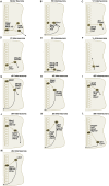
(A–M) Simplified schematic illustrations of development of MNs and ventral/dorsal subclass interneurons with important transcriptional factors.
FIGURE 5
FIGURE 5
The motor circuitry is shown in diagrammatic form in the lower panel. Here, neurons can be divided by projection patterns, that are ipsilateral, contralateral, or both. Three classes of neurotransmitter are found in the cord: excitatory glutamatergic (e.g., V2a), inhibitory GABAergic/glycinergic (e.g., V2b), and excitatory cholinergic neurons (e.g., motor neurons). Roles for neurons in defining rate (e.g., V1), left–right alternation (e.g., V0) and rhythmicity (e.g., V3), are emerging. Adapted from Alaynick et al. (2011).

Similar articles

Cited by

References

    1. Al-Mosawie A., Wilson J. M., Brownstone R. M. (2007). Heterogeneity of V2-derived interneurons in the adult mouse spinal cord. Eur. J. Neurosci. 26 3003–3015. 10.1111/j.1460-9568.2007.05907.x - DOI - PubMed
    1. Alaynick W. A., Jessell T. M., Pfaff S. L. (2011). SnapShot: spinal cord development. Cell 146 178–178. 10.1016/j.cell.2011.06.038 - DOI - PMC - PubMed
    1. Alexander G. M., Rogan S. C., Abbas A. I., Armbruster B. N., Pei Y., Allen A. J., et al. (2009). Remote control of neuronal activity in transgenic mice expressing evolved G protein-coupled receptors. Neuron 63 27–39. 10.1016/j.neuron.2009.06.014 - DOI - PMC - PubMed
    1. Alvarez F. J., Dewey D. E., McMillin P., Fyffe R. E. (1999). Distribution of cholinergic contacts on Renshaw cells in the rat spinal cord: a light microscopic study. J. Physiol. 515(Pt 3), 787–797. 10.1111/j.1469-7793.1999.787ab.x - DOI - PMC - PubMed
    1. Alvarez F. J., Jonas P. C., Sapir T., Hartley R., Berrocal M. C., Geiman E. J., et al. (2005). Postnatal phenotype and localization of spinal cord V1 derived interneurons. J. Comp. Neurol. 493 177–92. 10.1002/cne.20711 - DOI - PMC - PubMed