. 2015 Jun 16:6:126.
doi: 10.3389/fphar.2015.00126. eCollection 2015.

High mobility group box 1 contributes to wound healing induced by inhibition of dipeptidylpeptidase 4 in cultured keratinocytes

Affiliations

High mobility group box 1 contributes to wound healing induced by inhibition of dipeptidylpeptidase 4 in cultured keratinocytes

Tiziana Sinagra et al. Front Pharmacol. .

Abstract

Dipeptidyl peptidase 4 (DPP4) is expressed in various tissues, including the skin, and DPP4 inhibitors, that are currently used for the treatment of diabetes, may be effective also for complications of diabetes that affect the skin. To assess the role of DPP4 in keratinocytes, after creating a scratch wound in a monolayer of NTCC 2544 cells, we evaluated DPP4 expression and monitored wound repair over time, after treatment with the DPP4 inhibitor 1(((1-(hydroxymethyl)cyclopentyl)amino)acetyl)2,5-cis-pyrrolidinedicarbonitrile (DPP4-In). Expression of DPP4 increased early and was maintained up to 48 h following the scratch as shown by western blot and immunostaining. Treatment with 10 μM DPP4-In reduced DPP4 expression and significantly accelerated wound repair. This effect did not involve enhanced cell proliferation as shown by MTT proliferation assay, the lack of changes of cell cycle profiles and the slight inhibition of ERK phosphorylation. Enhancement of wound repair by DPP4 inhibition was prevented by the non-specific MMPs inhibitor GM6100 (5 μM). Treatment with DPP4-In increased the expression of high mobility group box 1 (HMGB1), a substrate of this enzyme, and exposure of NCTC 2544 cells to DPP4-In and exogenous HMGB1 (10 nM) produced a non-additive effect. Finally the healing promoting effect of DPP4-In was prevented by pretreatment with a neutralizing anti-HMGB1 antibody. The present results suggest that DPP4 inhibition contributes to enhanced wound healing by inducing keratinocytes to migrate into a scratched area. This effect seems to be independent of cell proliferation and involves enhanced production of HMGB1.

Keywords: HMGB1; diabetes; keratinocytes; skin.


Figures

FIGURE 1
FIGURE 1
Increase of DPP4 expression in NCTC 2544 keratinocytes subjected to scratch wound. DPP4 expression is analyzed under basal conditions (A) and following scratch wound (B,C). Expression of the enzyme appears upregulated already 8 h after scratch (B) as shown by immunostaining and its overexpression is maintained up to 48 h (C). In (A) immunostaining for DPP4 (upper panel) and a bright field image (lower panel) of a NCTC2544 monolayer (10x magnification). In (B,C) bright field and immunofluorescence images of the scratched area captured with a 10x (upper panels) and a 40x (lower panels) magnification. Western blot analysis confirms the overexpression at 24 and 48 h (D). Bars show densitometry vs α-tubulin and are mean +/– SE of at least three independent determinations. *p < 0.05 vs control as by one way ANOVA and Newman-Keuls test for significance.
FIGURE 2
FIGURE 2
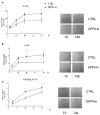
Inhibition of DPP4 increases wound closure in a monolayer of NTCC 2544 keratinocytes. Treatment with DPP4-In (10 μM) increases the ability of keratinocytes to migrate into the empty area and to repair the wound at all time points examined (A,B). Representative images of the wound repair over time are reported in (A). This effect is accompanied by reduction of the expression of DPP4 induced by scratch wound (scr), as by western blot analysis (C). Data are mean +/– SE of 3–5 independent experiments each run in quadruplicates. In (B), *p < 0.05 vs control by two-way ANOVA and Bonferroni post hoc test for significance; *p < 0.05 vs unscratched and **p < 0.05 vs untreated scratch (C) by one-way ANOVA and Newman-Keuls test for significance.
FIGURE 3
FIGURE 3
The healing promoting effect of DPP4-In does not involve increased cell proliferation. Treatment with increasing concentrations of DPP4-In does not modify cell proliferation as from MTT assay (A). Cell cycle analysis of NCTC 2544 cells treated with DPP4-In (10 μM) for 24 h does not modify the distribution of cells in different phases of cell cycle, as by flow cytometry after labeling of cells with propidium iodide. In (B), representative profiles of control (left panel) and DPP4-In (right panel) treated cells. Data summarized in the table are mean +/– SE of three independent experiments each run in quadruplicates. In (C), the increased phosphorylation of ERK induced by scratch wound is counteracted by DPP4-In (10 μM) treatment at 15 min and at 3 h. One representative plot is shown and expression of α-tubulin is reported for loading control. Data were analyzed by one-way ANOVA and Newman-Keuls test for significance. In (B), *p < 0.05 vs control and **p < 0.05 vs scratch alone.
FIGURE 4
FIGURE 4
DPP4-In requires MMPs to promote wound repair. The cell monolayer was scratched and pre-treated with the non-specific MMP inhibitor GM6001 (5 μM) for 30 min prior to addition of DPP4-In (10 μM) (A). In (B) representative blots of MMP2 and MMP9 24 h after scratch in the presence and absence of DPP4-In are shown. Data reported are the % of scratch repair at 24 h and represent mean +/– SE of three independent experiments. *p < 0.05 vs control and **p < 0.05 vs DPP4-In alone by one-way ANOVA and Newman-Keuls test.
FIGURE 5
FIGURE 5
Inhibition of DPP4 increases scratch repair in different cell types. Treatment with DPP4-In (10 μM) enhances wound repair in normal human keratinocytes (N-HEK) (A), keratinocytes from diabetic patients (D-HEK) (B) and fibroblasts from healthy human skin (C). In (A) and (B), DPP4-In was added at time 0 and scratch repair was monitored for the following 96 h. Data are mean +/– SE from three independent experiments each run in quadruplicates. *p < 0.05 by Two-way ANOVA and Bonferroni post hoc test for significance.
FIGURE 6
FIGURE 6
HMGB1 is involved in the healing promoting effect of DPP4-In. HMGB1 (10 nM) and SDF-1α (50 ng/ml) significantly increase wound repair over time in scratched NCTC 2544 keratinocytes (A). Co-treatment with DPP4-In (10 μM) and HMGB1 (10 nM) produces a non-additive effect (B), whereas co-addition of DPP4-In and SDF-1α (50 ng/ml) potentiates repair induced by DPP4-In alone (B). Western blot analysis shows that treatment with DPP4-In (10 μM, 24 h) counteracts the reduction of HMGB1, but not that of SDF-1α induced by scratch (C). In (D), treatment with a neutralizing anti-HMGB1 antibody (2.5 μg/ml) prevents the increase of scratch repair induced by treatment with DPP4-In (10 μM). Data are mean +/– SE of three to four independent experiments. In (A) and (B), *p < 0.05 vs control; §p < 0.05 vs DPP-In alone. In (C), *p < 0.05 vs control and **p < 0.05 vs scratch alone; in (D), *p < 0.05 vs control and **p < 0.05 vs DPP4-In alone. All data were analyzed by one-way ANOVA and Newman-Keuls test for significance.

References

    1. Avniel S., Arik Z., Maly A., Sagie A., Basst H. B., Yahana M. D., et al. (2006). Involvement of the CXCL12/CXCR4 pathway in the recovery of skin following burns. J. Invest. Dermatol. 126, 468–476. 10.1038/sj.jid.5700069 - DOI - PubMed
    1. Deacon C. F., Nauck M. A., Toft-Nielsen M., Pridal L., Willms B., Holst J. J. (1995). Both subcutaneously and intravenously administered glucagon-like peptide I are rapidly degraded from the NH2-terminus in type II diabetic patients and in healthy subjects. Diabetes 44, 1126–1131. 10.2337/diab.44.9.1126 - DOI - PubMed
    1. De Meester I., Vanhoof G., Hendriks D., Demuth H. U., Yaron A., Scharpé S. (1992). Characterization of dipeptidyl peptidase IV (CD26) from human lymphocytes. Clin. Chim. Acta 210, 23–34. 10.1016/0009-8981(92)90042-O - DOI - PubMed
    1. Falanga V. (2005). Wound healing and its impairment in the diabetic foot. Lancet 366, 1736–1743. 10.1016/S0140-6736(05)67700-8 - DOI - PubMed
    1. Fuchs H., Binder R., Greischel A. (2009). Tissue distribution of the novel DPP-4 inhibitor BI 1356 is dominated by saturable binding to its target in rats. Biopharm. Drug Dispos. 30, 229–240. 10.1002/bdd.662 - DOI - PubMed

LinkOut - more resources