

Automated Feature Extraction in Brain Tumor by Magnetic Resonance Imaging Using Gaussian Mixture Models
- PMID: 26136774
- PMCID: PMC4469084
- DOI: 10.1155/2015/868031
Automated Feature Extraction in Brain Tumor by Magnetic Resonance Imaging Using Gaussian Mixture Models
Erratum in
-
Corrigendum to "Automated Feature Extraction in Brain Tumor by Magnetic Resonance Imaging Using Gaussian Mixture Models".Int J Biomed Imaging. 2017;2017:3247974. doi: 10.1155/2017/3247974. Epub 2017 Aug 13. Int J Biomed Imaging. 2017. PMID: 28883830 Free PMC article.
Abstract
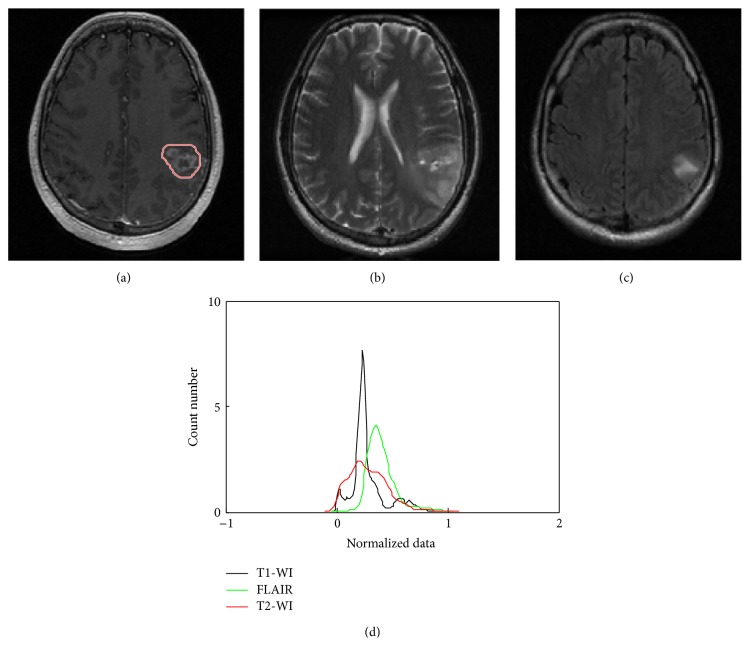
This paper presents a novel method for Glioblastoma (GBM) feature extraction based on Gaussian mixture model (GMM) features using MRI. We addressed the task of the new features to identify GBM using T1 and T2 weighted images (T1-WI, T2-WI) and Fluid-Attenuated Inversion Recovery (FLAIR) MR images. A pathologic area was detected using multithresholding segmentation with morphological operations of MR images. Multiclassifier techniques were considered to evaluate the performance of the feature based scheme in terms of its capability to discriminate GBM and normal tissue. GMM features demonstrated the best performance by the comparative study using principal component analysis (PCA) and wavelet based features. For the T1-WI, the accuracy performance was 97.05% (AUC = 92.73%) with 0.00% missed detection and 2.95% false alarm. In the T2-WI, the same accuracy (97.05%, AUC = 91.70%) value was achieved with 2.95% missed detection and 0.00% false alarm. In FLAIR mode the accuracy decreased to 94.11% (AUC = 95.85%) with 0.00% missed detection and 5.89% false alarm. These experimental results are promising to enhance the characteristics of heterogeneity and hence early treatment of GBM.
Figures


References
LinkOut - more resources
Full Text Sources
Other Literature Sources
