. 2015 Jun;9(6):2647-2650.
doi: 10.3892/ol.2015.3100. Epub 2015 Apr 3.

Prostate cancer incorrectly diagnosed as a rectal tumor: A case report

Affiliations

Prostate cancer incorrectly diagnosed as a rectal tumor: A case report

Zhi-Hua Liu et al. Oncol Lett. 2015 Jun.

Abstract

Colorectal cancer is the third most commonly diagnosed type of cancer in the world. Prostate adenocarcinoma is the most common male genitourinary tract malignancy, usually occurring after the age of 60. Prostate adenocarcinoma is a highly metastatic cancer. The common metastatic locations of prostate cancer are the bone, lung and liver. The elective locations are bones. Solitary rectal metastasis of prostate cancer is relatively rare. In the present study we report a case of solitary metastasis of a prostate adenocarcinoma with the prostatic capsule intact, which initially led to an incorrect diagnosis.

Keywords: prostate adenocarcinoma; rectal metastasis.


Figures
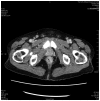
Figure 1.
Figure 1.
Transversal view of pelvic enhanced computed tomography evaluation of enhanced. Thickening of the low rectal wall is observed.
Figure 2.
Figure 2.
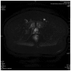
Transversal view of pelvic diffusion-weighted magnetic resonance imaging reveals low rectal cancer and a mass located between the prostate and fundus of the urinary bladder which was considered to be benign and derived from the prostate, while the prostate was well encapsulated.
Figure 3.
Figure 3.
Enteroscopy findings of the rectal tumor: a 3.5×3.5-cm in size, firm, poorly defined rectal protruding lesion with erosion of the surface, which was 3 cm away from the anus.
Figure 4.
Figure 4.
Microscopic findings of the rectal tumor (hematoxylin and eosin staining; magnification, ×400. Proliferation of atypical glandular cells was observed.

References

    1. Ito K. Prostate cancer in Asian men. Nat Rev Urol. 2014;11:197–212. doi: 10.1038/nrurol.2014.42. - DOI - PubMed
    1. Gu J, Chen N. Current status of rectal cancer treatment in China. Colorectal Dis. 2013;5:1345–1350. doi: 10.1111/codi.12269. - DOI - PubMed
    1. Reigman HI, Stokkel MP. Peripheral bone metastases in prostate cancer: a rare localization at initial presentation. Clin Nucl Med. 2004;29:335–336. doi: 10.1097/01.rlu.0000123327.62553.d0. - DOI - PubMed
    1. Nakamura T, Mohri H, Shimazaki M, et al. Esophageal metastasis from prostate cancer: diagnostic use of reverse transcriptase-polymerase chain reaction for prostate-specific antigen. J Gastroenterol. 1997;32:236–240. doi: 10.1007/BF02936374. - DOI - PubMed
    1. Larkin JO, Collins CG, Martin ST, et al. Paraesophageal lymph node metastasis from prostatic adenocarcinoma in a patient with esophageal squamous carcinoma. Dis Esophagus. 2005;18:124–126. doi: 10.1111/j.1442-2050.2005.00459.x. - DOI - PubMed

LinkOut - more resources