

ET-1 Plasma Levels, Aqueous Flare, and Choroidal Thickness in Patients with Retinitis Pigmentosa
- PMID: 26137317
- PMCID: PMC4468344
- DOI: 10.1155/2015/292615
ET-1 Plasma Levels, Aqueous Flare, and Choroidal Thickness in Patients with Retinitis Pigmentosa
Abstract
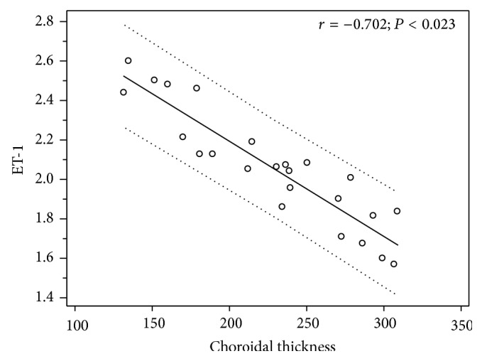
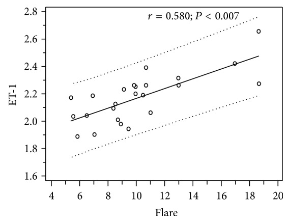
Purpose. To assess endothelin-1 (ET-1) plasma levels, choroidal thickness, and aqueous flare in patients with early stage retinitis pigmentosa (RP) and to search for possible correlations. Methods. We compared 24 RP patients with 24 healthy controls. Choroidal thickness and aqueous flare were measured, respectively, by using a spectral domain optical coherence tomography and a laser flare-cell meter, whereas plasma samples were obtained from each patient to evaluate ET-1 plasma levels. Results. Notably, RP subjects showed significantly increased ET-1 plasma levels and reduced choroidal thickness compared with controls: 2.143 ± 0.258 versus 1.219 ± 0.236 pg/mL, P < 0.002, and 226.75 ± 76.37 versus 303.9 ± 39.87 μm, P < 0.03, respectively. Higher aqueous flare values were also demonstrated in RP compared to controls: in detail, 10.51 ± 3.97 versus 5.66 ± 1.29 photon counts/ms, P < 0.0001. Spearman's correlation test highlighted that the increase of ET-1 plasma levels was related with the decrease of choroidal thickness (r = -0.702; P < 0.023) and the increase of aqueous flare (r = 0.580; P < 0.007). Conclusions. Early stage RP patients show a breakdown of blood-ocular barrier and increased ET-1 plasma levels and these findings may contribute to the reduction of choroidal thickness.
Figures

References
-
- Cellini M., Lodi R., Possati G. L., Sbrocca M., Pelle D., Giubilei N. Color doppler ultrasonography in Retinitis pigmentosa. Preliminary study. Journal Français d'Ophtalmologie. 1997;20(9):659–663. - PubMed
LinkOut - more resources
Full Text Sources
Other Literature Sources
