. 2015:2015:823139.
doi: 10.1155/2015/823139. Epub 2015 Jun 2.

A Novel Device to Exploit the Smartphone Camera for Fundus Photography

Affiliations

A Novel Device to Exploit the Smartphone Camera for Fundus Photography

Andrea Russo et al. J Ophthalmol. 2015.

Abstract

Purpose. To construct an inexpensive, convenient, and portable attachment for smartphones for the acquisition of still and live retinal images. Methods. A small optical device based on the principle of direct ophthalmoscopy was designed to be magnetically attached to a smartphone. Representative images of normal and pathological fundi were taken with the device. Results. A field-of-view up to ~20° was captured at a clinical resolution for each fundus image. The cross-polarization technique adopted in the optical design dramatically diminished corneal Purkinje reflections, making it possible to screen patients even through undilated pupils. Light emission proved to be well within safety limits. Conclusions. This optical attachment is a promising, inexpensive, and valuable alternative to the direct ophthalmoscope, potentially eliminating problems of poor exam skills and inexperienced observer bias. Its portability, together with the wireless connectivity of smartphones, presents a promising platform for screening and telemedicine in nonhospital settings. Translational Relevance. Smartphones have the potential to acquire retinal imaging for a portable ophthalmoscopy.


Figures
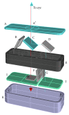
Figure 1
Figure 1
Exploded view of the D-Eye module (angles and distances between components are approximated). Retinal images are acquired using coaxial illumination and imaging paths thanks to a beam splitter (C). The blue arrow depicts the path of the light; red arrow depicts the path of fundus imaging. Device components are glass platelet (A) with imprinted negative lens (A′), photo-absorbing wall (B), beam splitter (C), mirror (D), plastic case (E), diaphragm (F), polarized filters (G, H), flash and camera glass (J, I), and magnetic external ring (K).
Figure 2
Figure 2
Picture of the prototypes magnetically attached to different iPhone (5 and 5s; Apple, Cupertino, CA) models.
Figure 3
Figure 3
The acquisition procedure is similar to traditional direct ophthalmoscopy, but the examiner can work from a comfortable position.
Figure 4
Figure 4
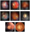
Representative retinal images taken with D-Eye. (a) Normal optic disc in an undilated child. (b) Normal posterior pole in a dilated 29-year-old woman. (c) Dry age-related maculopathy in an undilated 75-year-old man. (d) Optic nerve glioma in a 23-year-old undilated woman. (e) Posterior vitreous detachment in a dilated 72-year-old pseudophakic woman. (f) Waxy disc pallor and pigmentary changes in a 50-year-old man with retinitis pigmentosa. ((g) and (h)) Depiction of the same optic nerve head by D-Eye and Canon CR-2 Retinal Camera.
Figure 5
Figure 5
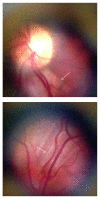
Cross-polarization-accentuated nerve-fiber layer definition (white arrows).

References

    1. Abramoff M. D., Garvin M. K., Sonka M. Retinal imaging and image analysis. IEEE Reviews in Biomedical Engineering. 2010;3:169–208. doi: 10.1109/RBME.2010.2084567. - DOI - PMC - PubMed
    1. Shields C. L., Materin M., Shields J. A. Panoramic imaging of the ocular fundus. Archives of Ophthalmology. 2003;121(11):1603–1607. doi: 10.1001/archopht.121.11.1603. - DOI - PubMed
    1. Ting D. S. W., Tay-Kearney M. L., Kanagasingam Y. Light and portable novel device for diabetic retinopathy screening. Clinical and Experimental Ophthalmology. 2012;40(1):e40–e46. doi: 10.1111/j.1442-9071.2011.02732.x. - DOI - PubMed
    1. Maamari R. N., Keenan J. D., Fletcher D. A., Margolis T. P. A mobile phone-based retinal camera for portable wide field imaging. British Journal of Ophthalmology. 2014;98(4):438–441. doi: 10.1136/bjophthalmol-2013-303797. - DOI - PubMed
    1. Tran K., Mendel T. A., Holbrook K. L., Yates P. A. Construction of an inexpensive, hand-held fundus camera through modification of a consumer ‘point-and-shoot’ camera. Investigative Ophthalmology and Visual Science. 2012;53(12):7600–7607. doi: 10.1167/iovs.12-10449. - DOI - PMC - PubMed