

Consensus nomenclature for CD8+ T cell phenotypes in cancer
- PMID: 26137416
- PMCID: PMC4485711
- DOI: 10.1080/2162402X.2014.998538
Consensus nomenclature for CD8+ T cell phenotypes in cancer
Abstract
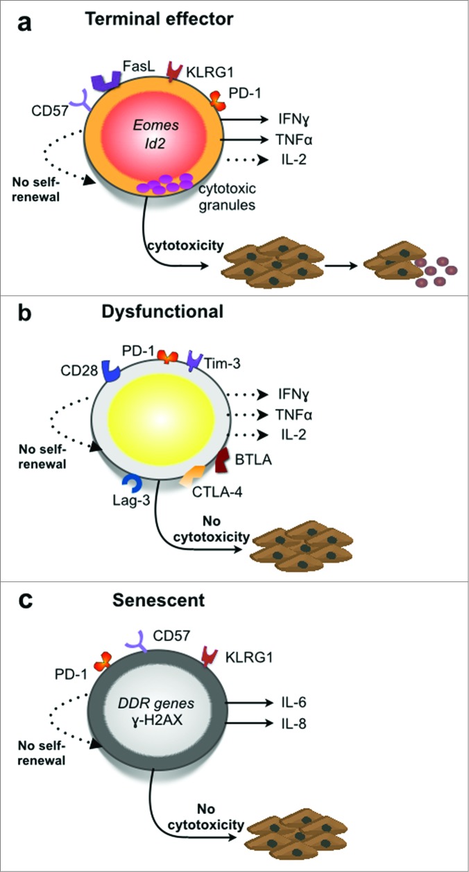
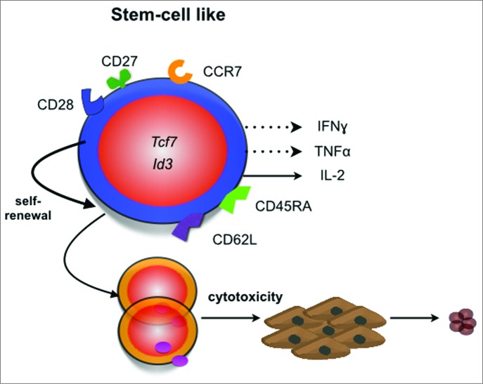
Whereas preclinical investigations and clinical studies have established that CD8+ T cells can profoundly affect cancer progression, the underlying mechanisms are still elusive. Challenging the prevalent view that the beneficial effect of CD8+ T cells in cancer is solely attributable to their cytotoxic activity, several reports have indicated that the ability of CD8+ T cells to promote tumor regression is dependent on their cytokine secretion profile and their ability to self-renew. Evidence has also shown that the tumor microenvironment can disarm CD8+ T cell immunity, leading to the emergence of dysfunctional CD8+ T cells. The existence of different types of CD8+ T cells in cancer calls for a more precise definition of the CD8+ T cell immune phenotypes in cancer and the abandonment of the generic terms "pro-tumor" and "antitumor." Based on recent studies investigating the functions of CD8+ T cells in cancer, we here propose some guidelines to precisely define the functional states of CD8+ T cells in cancer.
Keywords: CD8+ T cells; IFNγ; anergy; anticancer immunity; cytotoxicity; effector; exhaustion; senescence; stemness.
Figures
References
-
- Harty JT, Tvinnereim AR, White DW. CD8+ T cell effector mechanisms in resistance to infection. Ann Rev Immunol 2000; 18:275-308; PMID:; http://dx.doi.org/10.1146/annurev.immunol.18.1.275 - DOI - PubMed
-
- Uyttenhove C, Maryanski J, Boon T. Escape of mouse mastocytoma P815 after nearly complete rejection is due to antigen-loss variants rather than immunosuppression. J Exp Med 1983; 157:1040-52; PMID:; http://dx.doi.org/10.1084/jem.157.3.1040 - DOI - PMC - PubMed
-
- Nakayama E, Uenaka A. Effect of in vivo administration of Lyt antibodies. Lyt phenotype of T cells in lymphoid tissues and blocking of tumor rejection. J Exp Med 1985; 161:345-55; PMID:; http://dx.doi.org/10.1084/jem.161.2.345 - DOI - PMC - PubMed
-
- Shankaran V, Ikeda H, Bruce AT, White JM, Swanson PE, Old LJ, Schreiber RD. IFNgamma and lymphocytes prevent primary tumour development and shape tumour immunogenicity. Nature 2001; 410:1107-11; PMID:; http://dx.doi.org/10.1038/35074122 - DOI - PubMed
-
- Smyth MJ, Thia KY, Street SE, MacGregor D, Godfrey DI, Trapani JA. Perforin-mediated cytotoxicity is critical for surveillance of spontaneous lymphoma. J Exp Med 2000; 192:755-60; PMID:; http://dx.doi.org/10.1084/jem.192.5.755 - DOI - PMC - PubMed
Publication types
Grants and funding
LinkOut - more resources
Full Text Sources
Other Literature Sources
Research Materials