. 2014 Sep 22;1(1):72-9.
doi: 10.1016/j.ebiom.2014.09.001. eCollection 2014 Nov.

In Vivo Application of Tissue-Engineered Veins Using Autologous Peripheral Whole Blood: A Proof of Concept Study

Affiliations

In Vivo Application of Tissue-Engineered Veins Using Autologous Peripheral Whole Blood: A Proof of Concept Study

Michael Olausson et al. EBioMedicine. .

Abstract

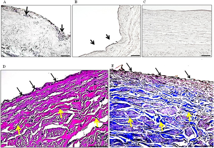
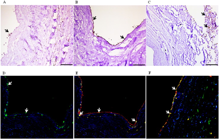
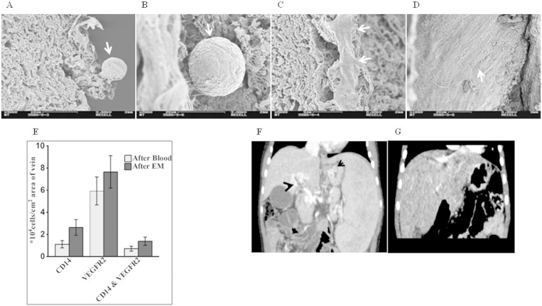
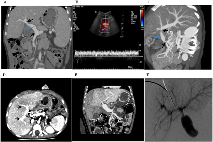
Vascular diseases are increasing health problems affecting > 25 million individuals in westernized societies. Such patients could benefit from transplantation of tissue-engineered vascular grafts using autologous cells. One challenge that has limited this development is the need for cell isolation, and risks associated with ex vivo expanded stem cells. Here we demonstrate a novel approach to generate transplantable vascular grafts using decellularized allogeneic vascular scaffolds, repopulated with peripheral whole blood (PWB) in vitro in a bioreactor. Circulating, VEGFR-2 +/CD45 + and a smaller fraction of VEGFR-2 +/CD14 + cells contributed to repopulation of the graft. SEM micrographs showed flat cells on the luminal surface of the grafts consistent with endothelial cells. For clinical validation, two autologous PWB tissue-engineered vein conduits were prepared and successfully used for by-pass procedures in two pediatric patients. These results provide a proof of principle for the generation of transplantable vascular grafts using a simple autologous blood sample, making it clinically feasible globally.

Keywords: Endothelial precursors; Tissue-engineering; Vascular diseases; Vein conduits.


Figures

Fig. 1
Fig. 1
A representative microscopic view of decellularized vein grafts. Masson's Trichrome staining of (A) decellularized vein (DV) showing no nuclei, indicating lack of endothelial and smooth muscle cells, but with abundant collagen still present (blue) and (B) normal vein showing presence of nuclei (black, arrows), cytoplasm (red/pink) and collagen (blue). (C) Negative controls. (D) Immunohistochemical staining of normal and DV for the various extracellular matrix components. Nuclei are seen only in normal but not in DV. In the DV although no cells/nuclei are seen, the ECM components stained positive indicating the retainment of important ECM proteins (n = 14). A–D: Scale bar = 75 μm.
Fig. 2
Fig. 2
Microscopic view of a representative bioengineered vein graft with peripheral whole blood. (A) Picture shows migration of cells (black) into the tissue after perfusion with blood, (B & C) presence of an endothelial cell monolayer on the luminal side after perfusion with endothelial cell medium and an overall improved architecture of the vein. Staining of tissue-engineered human veins recellularized with peripheral whole blood by (D) Verhoeff Van Gieson, showed presence of nuclei (black) for endothelial (black arrows) and smooth muscle cells (yellow arrows). The cytoplasm was stained pink and elastin black. (E) Masson's Trichrome staining showed presence of black nuclei (black), blue collagen and pink cytoplasm (n = 5). A–C: Scale bar = 75 μm, D & E: scale bar = 25 μm.
Fig. 3
Fig. 3
Phenotypic labeling of peripheral whole blood cells involved in repopulation of the bioengineered veins. (A) Cells staining positive (brown) for the tyrosine kinase receptor VEGFR-2 are found adhering to the lumen of the scaffold after perfusion of peripheral blood for 48 h. (B) These VEGFR-2 + cells formed a monolayer on the luminal side of the bioengineered vein after perfusion of endothelial cell medium for 4 days (brown). (C) Cells expressing the common leucocyte marker CD45 were found mainly in the media and adventitia (arrows). (D & E) A small fraction of cells in the lumen also stain positive for the monocyte marker CD14 (green), while double staining showed that some were also double positive (arrows) for VEGFR-2 (red). (F) Double staining showing that VEGFR-2 + cells were also positive for the endothelial marker CD31. Yellow color indicates co-localization of the two markers. The repopulating cells were negative for other markers such as CD3, CD19, CD68, CD133, CD34, CD56 and CD61 (n = 6).
Fig. 4
Fig. 4
Characterization of the recellularized veins and pre-transplant clinical data of the two patients. (A & B) Scanning electron micrographs of the recellularized veins showing binding of cells (arrows) to the lumen and in the media after perfusion with peripheral whole blood. (C & D) Further perfusion with endothelial cells medium resulted in formation of a continous smooth endothelial cell layer (arrows). (E) Higher numbers of VEGFR-2 + cells were found in the recellularized veins (n = 6) as compared to CD14 + cells both after perfusion with blood and endothelial cell medium (EM). (F & G) CT-angiographs of patients 1 & 2 respectively before transplantation. (F) Patient 1: showing varicose veins in the hilum (blue) and at the esophagus (black). (G) Patient 2: showing no intrahepatic portal system. In both cases, no portal circulation is observed.
Fig. 5
Fig. 5
Post-transplant clinical data for the two transplanted patients. (A) Patient 1: Postoperative CT-angiograph, showing the new graft (blue arrow) with good anastomotic sites (black arrows) and well established intrahepatic portal system. (B) The postoperative period was uneventful with good intrahepatic blood flows. The flow velocity showed 30–40 cm/s both in the portal vein and in the hepatic artery. (C) Patient 2: CT-angiography after first transplantation, showing a revascularized portal flow and intrahepatic portal system (blue arrow). At the six-month checkup, the patient had a reduced diameter at both anastomotic sites, (black arrows), however the intrahepatic portal vein system was significantly developed (D). Seven months later the patient received a second graft. (E) CT angiography day after surgery of the second graft revealed poor perfusion of the right portal system. (F) However, after a percutaneous angiography the right portal system was well perfused.

References

    1. Asahara T., Murohara T., Sullivan A. Isolation of putative progenitor endothelial cells for angiogenesis. Science. 1997;275(5302):964–967. - PubMed
    1. Bailey A.S., Willenbring H., Jiang S. Myeloid lineage progenitors give rise to vascular endothelium. Proc. Natl. Acad. Sci. U. S. A. 2006;103(35):13156–13161. - PMC - PubMed
    1. Bucala R., Spiegel L.A., Chesney J., Hogan M., Cerami A. Circulating fibrocytes define a new leukocyte subpopulation that mediates tissue repair. Mol. Med. 1994;1(1):71–81. - PMC - PubMed
    1. Clowes A.W., Kirkman T.R., Reidy M.A. Mechanisms of arterial graft healing. Rapid transmural capillary ingrowth provides a source of intimal endothelium and smooth muscle in porous PTFE prostheses. Am. J. Pathol. 1986;123(2):220–230. - PMC - PubMed
    1. Deutsch M., Meinhart J., Zilla P. Long-term experience in autologous in vitro endothelialization of infrainguinal ePTFE grafts. J. Vasc. Surg. 2009;49(2):352–362. (discussion 62) - PubMed