. 2015 Mar 28;2(5):467-75.
doi: 10.1016/j.ebiom.2015.03.020. eCollection 2015 May.

Allogeneic Mesenchymal Stem Cells Restore Endothelial Function in Heart Failure by Stimulating Endothelial Progenitor Cells

Affiliations

Allogeneic Mesenchymal Stem Cells Restore Endothelial Function in Heart Failure by Stimulating Endothelial Progenitor Cells

Courtney Premer et al. EBioMedicine. .

Abstract

Background: Endothelial dysfunction, characterized by diminished endothelial progenitor cell (EPC) function and flow-mediated vasodilation (FMD), is a clinically significant feature of heart failure (HF). Mesenchymal stem cells (MSCs), which have pro-angiogenic properties, have the potential to restore endothelial function. Accordingly, we tested the hypothesis that MSCs increase EPC function and restore flow-mediated vasodilation (FMD).

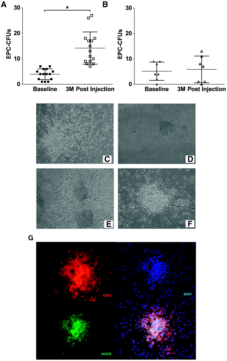
Methods: Idiopathic dilated and ischemic cardiomyopathy patients were randomly assigned to receive autologous (n = 7) or allogeneic (n = 15) MSCs. We assessed EPC-colony forming units (EPC-CFUs), FMD, and circulating levels of vascular endothelial growth factor (VEGF) in patients before and three months after MSC transendocardial injection (n = 22) and in healthy controls (n = 10).

Findings: EPC-colony forming units (CFUs) were markedly reduced in HF compared to healthy controls (4 ± 3 vs. 25 ± 16 CFUs, P < 0.0001). Similarly, FMD% was impaired in HF (5.6 ± 3.2% vs. 9.0 ± 3.3%, P = 0.01). Allogeneic, but not autologous, MSCs improved endothelial function three months after treatment (Δ10 ± 5 vs. Δ1 ± 3 CFUs, P = 0.0067; Δ3.7 ± 3% vs. Δ-0.46 ± 3% FMD, P = 0.005). Patients who received allogeneic MSCs had a reduction in serum VEGF levels three months after treatment, while patients who received autologous MSCs had an increase (P = 0.0012), and these changes correlated with the change in EPC-CFUs (P < 0.0001). Lastly, human umbilical vein endothelial cells (HUVECs) with impaired vasculogenesis due to pharmacologic nitric oxide synthase inhibition, were rescued by allogeneic MSC conditioned medium (P = 0.006).

Interpretation: These findings reveal a novel mechanism whereby allogeneic, but not autologous, MSC administration results in the proliferation of functional EPCs and improvement in vascular reactivity, which in turn restores endothelial function towards normal in patients with HF. These findings have significant clinical and biological implications for the use of MSCs in HF and other disorders associated with endothelial dysfunction.

Keywords: Autografts; Nitric oxide; Regenerative medicine; Vascular endothelium-dependent relaxation; Vasculogenesis.


Figures

Fig. 1
Fig. 1
Endothelial function in patients with heart failure, including dilated and ischemic cardiomyopathy. (A) Patients (n = 22) have impaired endothelial progenitor cell-colony forming units (EPC-CFUs) compared to healthy controls (n = 10, *P < 0.0001, t test). (B) Patients (n = 22) have reduced flow-mediated vasodilation (FMD%) compared to healthy controls (n = 10, P = 0. 01, t-test).
Fig. 2
Fig. 2
Endothelial colony forming units in heart failure patients treated with either allogeneic or autologous mesenchymal stem cells (MSCs). (A) Patients treated with allogeneic MSCs had a significant improvement in endothelial progenitor cell-colony forming units (EPC-CFUs) 3 months post-treatment (n = 15, *P < 0. 0001, t-test). (B) Patients treated with autologous MSCs had no change in EPC-CFUs post-treatment (n = 7, P = NS, t-test). (C–F) Representative EPC-CFUs plated on fibronectin for 5 days before (C, D) and after (E, F) MSC administration (magnification 20 ×). (G) Colonies are positive for the endothelial cell markers CD31 (red) and VEGFR (green) (magnification 20 ×).
Fig. 3
Fig. 3
Flow-mediated vasodilation (FMD) measurements before and after mesenchymal stem cell (MSC) treatment. (A) Patients treated with allogeneic MSCs had an increase in FMD% 3 months post-injection (n = 15, *P = 0.0002, t-test). (B) Patients treated with autologous MSCs had no significant difference in FMD% 3 months post-injection (n = 7, P = NS, t-test). (C) There is a strong correlation between the absolute change in FMD% and the absolute change in endothelial progenitor cell-colony forming units (EPC-CFUs) from baseline to 3 months post-MSC injection in all patients (*P = 0.0004, R = 0.68, Pearson correlation).
Fig. 4
Fig. 4
Serum vascular endothelium growth factor (VEGF) concentration in patients and controls. (A) Patients (n = 14) have a higher level of circulating VEGF compared to controls (n = 9) at baseline (*P = 0.0009, t-test; within-group, P = 0.21, P < 0.001, respectively, D'Agostino-Pearson omnibus normality test). (B) Patients who received allogeneic MSCs (n = 9) had a decrease in VEGF serum levels post-injection (Δ-547.5 ± 350.8 pg/mL, P = 0.0015, t-test; within-group P = 0.96, D'Agostino-Pearson omnibus normality test), while patients who received autologous MSCs (n = 5) had an increase in serum VEGF post-injection (Δ814.1 ± 875.8), and there was a difference between the groups (P = 0.0012, t-test). (C) There is a correlation between endothelial progenitor cell-colony forming units (EPC-CFUs) and serum VEGF in patients at both baseline and 3 months post-MSC treatment (R = − 0.421, P = 0.026, Pearson correlation). (D) The change in EPC-CFUs from baseline to 3 months post-treatment strongly correlated with the change in VEGF (R = − 0.863,*P < 0.0001, Pearson correlation).
Fig. 5
Fig. 5
Effect of autologous and allogeneic mesenchymal stem cell (MSC) treatment on vasculogenesis. (A–D) Representative pictures of human umbilical vein endothelial cells (HUVECs) alone (n = 3), HUVECs with the nitric oxide synthase inhibitor, L-NG-Nitroarginine methyl ester (L-NAME) (n = 3), HUVECs with L-NAME and allogeneic MSC-conditioned media (CM) (n = 5), and HUVECs with L-NAME and autologous MSC-CM (n = 7) after 6 h on Matrigel (magnification 10 ×). (E) L-NAME greatly reduced vascular index (*P < 0.05, ANOVA), and only allogeneic-CM restored it (*P < 0.05, ANOVA).

Comment in

  • Mesenchymal Stem Cells & Endothelial Function.
    Fish KM, Hajjar RJ. Fish KM, et al. EBioMedicine. 2015 Apr 29;2(5):376-7. doi: 10.1016/j.ebiom.2015.04.015. eCollection 2015 May. EBioMedicine. 2015. PMID: 26137582 Free PMC article. No abstract available.

References

    1. Asumda F.Z. Age-associated changes in the ecological niche: implications for mesenchymal stem cell aging. Stem Cell Res. Ther. 2013;4(3):47. - PMC - PubMed
    1. Blum A. Heart failure—new insights. Isr. Med. Assoc. J. 2009;11(2):105–111. - PubMed
    1. Bui A.L., Horwich T.B., Fonarow G.C. Epidemiology and risk profile of heart failure. Nat. Rev. Cardiol. 2011;8(1):30–41. - PMC - PubMed
    1. Cao Y., Gomes S., Rangel E.B. S-nitrosoglutathione reductase-dependent PPARγ denitrosylation participates in adipogenesis and osteogenesis. J. Clin. Investig. 2015;125(4) - PMC - PubMed
    1. Chen L., Tredget E.E., Wu P.Y., Wu Y. Paracrine factors of mesenchymal stem cells recruit macrophages and endothelial lineage cells and enhance wound healing. PLoS ONE. 2008;3(4):e1886. - PMC - PubMed

MeSH terms

Substances