. 2015 Aug 14;117(5):424-36.
doi: 10.1161/CIRCRESAHA.114.305393. Epub 2015 Jul 2.

Angiotensin II Induces Skeletal Muscle Atrophy by Activating TFEB-Mediated MuRF1 Expression

Affiliations

Angiotensin II Induces Skeletal Muscle Atrophy by Activating TFEB-Mediated MuRF1 Expression

Philipp Du Bois et al. Circ Res. .

Abstract

Rationale: Skeletal muscle wasting with accompanying cachexia is a life threatening complication in congestive heart failure. The molecular mechanisms are imperfectly understood, although an activated renin-angiotensin aldosterone system has been implicated. Angiotensin (Ang) II induces skeletal muscle atrophy in part by increased muscle-enriched E3 ubiquitin ligase muscle RING-finger-1 (MuRF1) expression, which may involve protein kinase D1 (PKD1).

Objective: To elucidate the molecular mechanism of Ang II-induced skeletal muscle wasting.

Methods and results: A cDNA expression screen identified the lysosomal hydrolase-coordinating transcription factor EB (TFEB) as novel regulator of the human MuRF1 promoter. TFEB played a key role in regulating Ang II-induced skeletal muscle atrophy by transcriptional control of MuRF1 via conserved E-box elements. Inhibiting TFEB with small interfering RNA prevented Ang II-induced MuRF1 expression and atrophy. The histone deacetylase-5 (HDAC5), which was directly bound to and colocalized with TFEB, inhibited TFEB-induced MuRF1 expression. The inhibition of TFEB by HDAC5 was reversed by PKD1, which was associated with HDAC5 and mediated its nuclear export. Mice lacking PKD1 in skeletal myocytes were resistant to Ang II-induced muscle wasting.

Conclusion: We propose that elevated Ang II serum concentrations, as occur in patients with congestive heart failure, could activate the PKD1/HDAC5/TFEB/MuRF1 pathway to induce skeletal muscle wasting.

Keywords: angiotensin II; gene expression regulation; heart failure; histone deacetylase 5; muscle RING-finger-1; protein kinase D; transcription factor EB.


Conflict of interest statement

The authors are not aware of interest conflicts.

Figures

Figure 1
Figure 1. A cDNA expression screen identified the transcription factor EB (TFEB) as activator of the human MuRF1 promoter
(A) Luciferase assays performed on cell extracts of COS-7 cells transfected with Hs_MuRF1-Luc (−543 bp) with increasing amounts of FLAG-TFEB (TFEB) or empty (−) plasmid. Luciferase activity was normalized to expression of CMV-LacZ and expressed as fold-increase. Data are represented as mean ± SD. **P<0.005, ***P<0.001. (B) qRT-PCR analysis of TFEB expression in skeletal muscle, heart, liver and spleen of C57Bl6/N mice (n = 4–5). GAPDH expression was used as reference. Data are represented as mean ± SD. GP indicates gastrocnemius/plantaris, Sol, soleus; TA, tibialis anterior, EDL, extensor digitorum longus; LA, left atrium; RA, right atrium; LV, left ventricle; RV, right ventricle. (C) qRT-PCR of MuRF1, MuRF2 and MuRF3 expression following transfection of cDNA expression plasmids encoding for FLAG-TFEB (+, TFEB) or empty (−) expression plasmid in C2C12 myoblasts for 24 h. GAPDH expression was used as reference. Data are represented as mean ± SD. **P<0.005. n = 3 each. (D) C2C12 cells were transfected with expression plasmids encoding wild type FLAG-TFEB (TFEB) or empty (control) expression plasmid for 24h, as indicated. Immunoblotting (IB) with anti-FLAG, anti-MuRF1 or anti-GAPDH antibody was performed. (E, F) C2C12 myoblasts were transfected with scrambled control siRNA (siScr, −) or siRNA targeting TFEB (siTFEB, +, 100 nM each) for 24h. (E) qRT-PCR analysis was performed to measure TFEB and MuRF1 expression. GAPDH expression was used as reference. Data are represented as mean ± SD. *P<0.05, ***P<0.001. n = 3 each. (F) Western blot analysis was performed with anti-TFEB, anti-MuRF1 or anti-GAPDH antibody.
Figure 2
Figure 2. TFEB regulates MuRF1 expression via conserved E-box elements
(A) COS-7 cells were transfected with wild type FLAG-TFEB expression plasmid or empty vector control (control), along with MuRF1-promoter constructs, as indicated. Data are represented as mean ± SD. *P<0.05, **P<0.01, ***P<0.005. (B) Schematic diagram of the human MuRF1-promoter. Positions of conserved E-box motifs (CANNTG) in the MuRF1-promoter relative to the transcription start (ATG) are indicated. Alignment shows genomic homology of individual E-box motifs between indicated species. Homo sapiens (mut.) indicates mutated nucleotides to inactivate individual E-boxes (mutated nucleotides are shown in bold). * indicates homology. (C) COS-7 cells were transfected with a TFEB expression plasmid and the indicated MuRF1-promoter constructs (−543 bp) harboring E-box mutations as shown in (B). Data are represented as mean ± SD. *P<0.05, ***P<0.005. (D) Chromatin immunoprecipitation (ChIP) assay performed on chromatin from C2C12 myoblasts using antibodies against TFEB. Primers flanking E-boxes 1, 2 and 3 of the MuRF1-promoter were used. Values indicate the fold-enrichment over chromatin immunoprecipitated with antibodies against IgG. n=5. (E, F) ChIP assay performed on chromatin from angiotensin II (Ang II, +) and vehicle treated (−) C2C12 myoblasts (E). ChIP assay performed on chromatin from serum starved (+) and untreated (−) C2C12 myoblasts (F). Chromatin was immunoprecipitated with antibodies against TFEB. Antibodies against IgG were used as control. n=3.
Figure 3
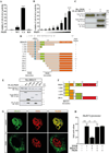
Figure 3. Induction of MuRF1 expression is mediated through TFEB’s bHLH domain
(A) Schemes of wild type TFEB and its deletion mutants used in this study. The presence of wild type and mutant TFEB protein in the nucleus (N) or cytoplasm (C), as detected by immunostaining, their ability to induce MuRF1 expression in luciferase assays, or their interaction with HDAC5 in coimmunoprecipitation assays, respectively, are indicated. GR indicates glycine-rich domain; bHLH, basic Helix-Loop-Helix domain; LZ, Leucine Zipper domain; PR, Proline-rich domain. (B) The subcellular distribution of wild type FLAG-TFEB and its deletion mutants in transfected C2C12 myoblasts was detected by immunofluorescence using anti-TFEB antibody together with A488-coupled secondary antibody. Nuclei were stained with DAPI (blue); scale bar 20 µm. (C) HEK293 cells were transfected with the Hs_MuRF1_Luc reporter construct (−543 bp) and expression plasmids encoding wild type or mutant TFEB, as indicated. Luciferase activity was normalized to expression of CMV-LacZ and calculated as fold-increase. Data are represented as mean ± SD.*P<0.05; ***P<0.001. n = 3. (D) C2C12 cells were transfected with expression plasmids encoding wild type (WT) FLAG-TFEB, TFEB deletion mutant ∆299–352 or empty expression plasmid (control) for 24 h, as indicated. Immunoblotting (IB) with anti-FLAG, anti-MuRF1 or anti-GAPDH antibody was performed.
Figure 4
Figure 4. TFEB induced MuRF1 expression is inhibited by HDAC5
(A) HEK293 cells were transfected with MuRF1-luciferase reporter (−543 bp) and expression plasmids encoding wild type TFEB, histone deacetylase (HDAC) 5 or empty vector control as indicated. Data are represented as mean ± SD. ***P<0.001. n = 3. (B) The subcellular distribution of wild type TFEB-GFP and HDAC5-Myc in transfected C2C12 myoblasts was detected by immunofluorescence. (a + b) indicate augmentations. neg. = negative control. (C) Co-IP with lysates from HEK293 cells expressing Myc-HDAC5 and FLAG-TFEB (top panel), or TFEB-Myc(His)6 and FLAG-HDAC5 (bottom panel), as indicated. Extracts were immunoprecipitated (IP) with anti-FLAG antibody and detected with an antibody directed against Myc. Input proteins were detected by Western blot (IB) with anti-FLAG and anti-Myc antibodies, respectively. n = 5. (D) Co-IP assay with lysates from HEK293 cells expressing wild type FLAG-TFEB or TFEB deletion mutants along with wild type Myc-HDAC5, as indicated. IP of TFEB-fusion proteins were using anti-FLAG antibody and detection by anti-Myc antibody. Input proteins were detected by Western blot (IB) with antibodies directed against FLAG and Myc. n = 3. (E) Co-IP assay with lysates from HEK293 cells expressing wild type Myc-HDAC5 or HDAC5 deletion mutants along with wild type FLAG-TFEB, as indicated. TFEB-fusion proteins were immunoprecipitated (IP) with anti-FLAG antibody and detected with an antibody directed against Myc. Input proteins were detected by immunoblot (IB) with antibodies directed against FLAG and Myc. n = 3. (F) HEK293 cells were transfected with the Hs_MuRF1_Luc reporter construct (−543 bp) and expression plasmids encoding wild type TFEB, TFEB deletion mutants and wild type HDAC5 as indicated. Data are represented as mean ± SD. **P<0.01, ***P<0.005. n = 3. (G) HEK293 cells were transfected with the Hs_MuRF1_Luc reporter construct (−543 bp) and expression plasmids encoding wild type TFEB, wild type HDAC5 and HDAC5 deletion mutants. Data are represented as mean ± SD. **P<0.01, ***P<0.005. n = 3.
Figure 5
Figure 5. PKD1 relieves HDAC5-mediated TFEB repression
(A) HEK293 cells were transfected with UAS-luciferase and expression plasmids encoding GAL4 fused to the wild type HDAC5 N-terminal extension together with 14-3-3-VP16 and expression plasmids of wild type (WT), constitutive active (CA) or kinase inactive (KD) PKD1 as indicated. Values were normalized to expression of CMV-LacZ and calculated as fold-increase. Data are represented as mean ± SD. *P<0.05; ** P<0.005. n = 3. (B) HEK293 cells were transfected with UAS-luciferase and expression plasmids encoding GAL4 fused to the wild type HDAC5 N-terminal extension together with 14-3-3-VP16 and increasing amounts of expression plasmids of PKD1 CA (from 6.25 ng to 400 ng), as indicated. Values were normalized to expression of CMV-LacZ and calculated as fold-increase. Data are represented as mean ± SD. *P<0.05, **P<0.01, ***P<0.005. n = 3. (C) Coimmunoprecipitation assay with lysates from COS-7 cells expressing Myc-PKD1 and FLAG-HDAC5, as indicated. HDAC5-fusion proteins were immunoprecipitated (IP) with anti-FLAG antibody and PKD1-fusion proteins were detected with an antibody directed against Myc. Input proteins were detected by Western blot (IB) with antibodies directed against the FLAG or Myc tag. n=3. (D) Based on the coimmunoprecipitation data, amino acids 360 to 601 of HDAC5 were identified to be required for physical interaction with PKD1 and, therefore, define a PKD1 binding site. (E) Coimmunoprecipitation assay of FLAG-PKD1 deletion mutants coexpressed with Myc-HDAC5 to identify the HDAC5 binding domain of PKD1. PKD1-fusion proteins were immunoprecipitated (IP) with anti-FLAG antibody and HDAC5-fusion proteins were detected with an antibody directed against Myc. Input proteins were detected by Western blot (IB) with antibodies directed against the FLAG or Myc tag. n = 3. (F) Based on the coimmunoprecipitation data, amino acids 1 to 201 of PKD1 were identified to be required for physical interaction with HDAC5 and, therefore, define a HDAC5 docking site. Positions of cysteine rich region 1a (C1a, yellow), C1b (orange), pleckstrin homology domain (PH; green) and kinase domain (red) of PKD1 are depicted. (G) COS-7 cells were transfected with GFP-HDAC5 and FLAG-TFEB together with an empty vector (pcDNA_3.1) or constitutive active PKD1. Immunostaining was performed with anti-FLAG and anti-GFP antibody. (H) HEK293 cells were transfected with expression plasmids encoding wild type FLAG-TFEB, HDAC5-Myc, or PKD1 CA proteins, as indicated, together with the Hs_MuRF1_Luc reporter construct (−543 bp). Values were normalized to expression of CMV-LacZ and calculated as the fold-increase in luciferase to CMV-LacZ ratio compared to the reporter alone. Data are represented as mean ± SD. *P<0.05; ** P<0.005. n = 5.
Figure 6
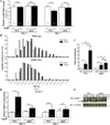
Figure 6. TFEB knockdown reduces endogenous MuRF1 expression and inhibits Ang II induced atrophy of C2C12 myocytes
(A) qRT-PCR analysis of MuRF1 expression in C2C12 myotubes following transfection with scrambled control siRNA (siScr) and siRNA targeting TFEB (siTFEB; 100 nM each) for 24h and following treatment with 500 nM Ang II or vehicle (−) for 24 h. GAPDH expression was used as reference. Data are represented as mean ± SEM. **P<0.01. n = 3. (B) qRT-PCR analysis of MuRF1 expression in C2C12 myoblasts following transfection with signal-resistant FLAG-HDAC5 S259/498A (harboring alanines in place of serines 259 and 498 which are required for nuclear export of HDAC5). GAPDH expression was used as reference. Data are represented as mean ± SEM. *P<0.05. n = 3. (C) C2C12 myoblasts were transfected with scrambled control siRNA (siScr) or siRNA targeting TFEB (siTFEB; 100 nM each), differentiated for 5 days, and treated with 500 nM Ang II or vehicle for 48h. Myotubes were photographed and myotube width was measured using ImageJ software. Number of myotubes belonging to a given range of myotube diameters is shown in a size distribution diagram. (D) Changes in mean myotube width following siRNA mediated knockdown of TFEB and Ang II or vehicle treatment is shown. *P<0.05, **P<0.01, ***P<0.005. n = 3 each.
Figure 7
Figure 7. Ang II induced skeletal muscle atrophy is attenuated in PKD1 cKO mice
Ang II (1.5 µg/KG/min) was delivered chronically to 8- to 10-wk-old male PKD1 wild type (WT; PKD1WT/WT; MCK-CRE) and skeletal muscle loss of function PKD1loxP/loxP; MCK-CRE (cKO) mice for 24h and 7 days, respectively, by using an implanted osmotic minipump. Vehicle treated wild type (PKD1WT/WT; MCK-CRE) and cKO mice were used as controls. Numbers of animals used are indicated. (A) Gastrocnemius/plantaris to tibia length ratios are shown. **P<0.01. n.s. not significant. (B) Myocyte cross sectional area (MCSA) of histological sections from gastrocnemius/plantaris (±SEM) of WT and cKO mice after 7 days of Ang II or vehicle treatment. Number of myotubes belonging to the given range of MCSA are shown in a size distribution diagram. (C) Chromatin immunoprecipitation (ChIP) assay performed on chromatin from gastrocnemius/plantaris of 24h Ang II and vehicle treated WT and cKO mice using anti-TFEB antibody. Primers flanking E-box 1 of the MuRF1 promoter were used. Values indicate the fold-enrichment over chromatin immunoprecipitated with anti-IgG antibody. n=3. *P<0.05, **P<0.01, n.s. not significant. (D) MuRF1 expression in gastrocnemius/plantaris (±SEM) of WT and cKO mice after 24h and 7 days of Ang II or vehicle treatment. GAPDH was used as a reference. Data are presented as mean±SEM. *P<0.05. (E) Western blot analysis on protein lysates from gastrocnemius/plantaris of 24h Ang II and vehicle treated WT and cKO mice using anti-MuRF1 and anti-GAPDH antibody. n.s., no specific signal.
Figure 8
Figure 8. The Ang II/PKD1/HDAC5 signaling pathway regulates TFEB-induced MuRF1 expression
The PKD1/HDAC5/TFEB/MuRF1 axis mediates Ang II induced skeletal muscle atrophy. Nuclear TFEB specifically binds to conserved E-box motifs in the MuRF1 promoter localized close to its transcription start site. PKD1 together with HDAC5 controls TFEB activity at the MuRF1 promoter. Inhibition of this signaling pathway could be important to combat Ang II associated muscle wasting disorders such as cardiac cachexia.

References

    1. Ciciliot S, Rossi AC, Dyar KA, Blaauw B, Schiaffino S. Muscle type and fiber type specificity in muscle wasting. Int J Biochem Cell Biol. 2013;45:2191–2199. - PubMed
    1. Borina E, Pellegrino MA, D'Antona G, Bottinelli R. Myosin and actin content of human skeletal muscle fibers following 35 days bed rest. Scandinavian journal of medicine & science in sports. 2010;20:65–73. - PubMed
    1. Snow LM, Sanchez OA, McLoon LK, Serfass RC, Thompson LV. Effect of endurance exercise on myosin heavy chain isoform expression in diabetic rats with peripheral neuropathy. American journal of physical medicine & rehabilitation / Association of Academic Physiatrists. 2005;84:770–779. - PubMed
    1. Ciechanover A. The ubiquitin proteolytic system: From a vague idea, through basic mechanisms, and onto human diseases and drug targeting. Neurology. 2006;66:S7–S19. - PubMed
    1. Vaughan VC, Martin P, Lewandowski PA. Cancer cachexia: Impact, mechanisms and emerging treatments. J Cachexia Sarcopenia Muscle. 2013;4:95–109. - PMC - PubMed

Publication types

MeSH terms

Substances