. 2015 Jul 3:6:7520.
doi: 10.1038/ncomms8520.

Impaired protein translation in Drosophila models for Charcot-Marie-Tooth neuropathy caused by mutant tRNA synthetases

Affiliations

Impaired protein translation in Drosophila models for Charcot-Marie-Tooth neuropathy caused by mutant tRNA synthetases

Sven Niehues et al. Nat Commun. .

Erratum in

Abstract

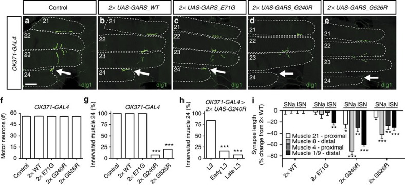
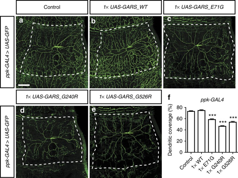
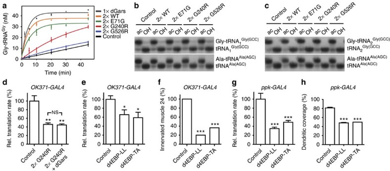
Dominant mutations in five tRNA synthetases cause Charcot-Marie-Tooth (CMT) neuropathy, suggesting that altered aminoacylation function underlies the disease. However, previous studies showed that loss of aminoacylation activity is not required to cause CMT. Here we present a Drosophila model for CMT with mutations in glycyl-tRNA synthetase (GARS). Expression of three CMT-mutant GARS proteins induces defects in motor performance and motor and sensory neuron morphology, and shortens lifespan. Mutant GARS proteins display normal subcellular localization but markedly reduce global protein synthesis in motor and sensory neurons, or when ubiquitously expressed in adults, as revealed by FUNCAT and BONCAT. Translational slowdown is not attributable to altered tRNA(Gly) aminoacylation, and cannot be rescued by Drosophila Gars overexpression, indicating a gain-of-toxic-function mechanism. Expression of CMT-mutant tyrosyl-tRNA synthetase also impairs translation, suggesting a common pathogenic mechanism. Finally, genetic reduction of translation is sufficient to induce CMT-like phenotypes, indicating a causal contribution of translational slowdown to CMT.


Figures

Figure 1
Figure 1. CMT-mutant GARS expression shortens lifespan and induces motor performance deficits.
(a) Schematic representation of the GARS protein, with CMT-associated mutations and functional domains indicated. Mutations in blue result in loss of aminoacylation activity, mutations in green do not affect enzymatic activity and mutations in black have not been evaluated. Mutations modelled in this study are framed. (b) Kaplan–Meier survival curves displaying the lifespan of female flies ubiquitously expressing two copies of GARS transgenes from the adult stage onwards. N=182–270. (c) Bar graph displaying average climbing speed in a negative geotaxis assay of female flies expressing GARS in motor neurons (OK371-GAL4). OK371-GAL4>GARS_G240R and OK371-GAL4>2 × GARS_G526R flies displayed developmental lethality and could not be assessed. N=100. Error bars represent s.e.m. *P<0.05; ***P<1 × 10−37.
Figure 2
Figure 2. Selective mutant GARS expression in motor neurons induces progressive muscle denervation in a proximo–distal gradient.
(ae) NMJs of third instar larvae expressing GARS in motor neurons (OK371-GAL4) were visualized by staining for the postsynaptic marker discs large 1 (dlg1). Arrows indicate the NMJ on muscle 24, which is missing in the vast majority of GARS_G240R and GARS_G526R animals. Scale bar, 50 μm. (f) No differences in motor neuron numbers were found between control and GARS larvae; one-way analysis of variance (ANOVA) with Bonferroni correction; P=0.88; N=21–33. (g) Quantification of the percentage of animals with muscle 24 innervated; χ2-Test; ***P<1 × 10⁻⁸; N=26. (h) Muscle 24 innervation in OK371-GAL4>2 × GARS_G240R larvae at different developmental stages (L2: second instar and L3: third instar). N=51, 42 and 30 NMJs; Mann–Whitney U-test; ***P<0.005 versus L2. (i) The effect of motor neuron-selective expression of GARS transgenes on synapse length on muscles 21, 8, 4 and 1/9 was quantified and plotted as the percentage change from 2 × GARS_WT. Muscles 8 and 21 are both innervated by the SNa motor nerve, whereas muscles 4 and 1/9 are innervated by the ISN motor nerve. Phenotypic strength ranged from G240R>G526R>E71G, and for all mutants, the phenotypic severity showed a proximo–distal gradient, with distal muscle (8 and 1/9) more severely affected than proximal ones (21 and 4); one-way ANOVA with Bonferroni correction; *P<0.05, **P<0.01, ***P<0.00005; N=12–14. Error bars represent s.e.m.
Figure 3
Figure 3. Selective mutant GARS expression in class IV multidendritic sensory neurons induces dendritic morphology defects.
(ae) Class IV multidendritic sensory neurons in the third instar larval body wall were visualized by ppk-GAL4-driven mCD8-GFP with or without co-expression of GARS. The dendritic tree of individual neurons is delineated. Scale bar, 100 μm. (f) Quantification of the percentage of dendritic coverage; one-way analysis of variance with Bonferroni correction; ***P<1 × 10⁻⁹; N=15–20. Error bars represent s.e.m.
Figure 4
Figure 4. Mutant and WT GARS and YARS proteins display similar subcellular localization in motor and sensory neurons.
(ah) GARS immunostaining reveals the subcellular localization of GARS proteins at third instar larval NMJs (ad) and motor neuron cell bodies (eh) upon expression in motor neurons (OK371-GAL4). (it) Subcellular localization of YARS proteins at third instar larval NMJs (il; nsyb-GAL4), motor neuron cell bodies (mp; nsyb-GAL4) and class IV multidendritic sensory neurons (qt; ppk-GAL4), as revealed by YARS immunostaining. Scale bars, 20 μm (ap) and 100 μm (qt).
Figure 5
Figure 5. Mutant GARS and YARS expression reduces global protein translation rates in motor and sensory neurons in vivo.
(ad) FUNCAT labelling in motor neurons of larvae co-expressing dMetRSL262G-EGFP and two copies of GARS transgenes (OK371-GAL4). Scale bar, 10 μm. (e) Quantification of FUNCAT signal intensity revealed ∼60% reduction of translation rates in motor neurons expressing GARS_G240R and GARS_G526R. Average±s.e.m. relative to GARS_WT (100%); Mann–Whitney U-test; ***P<0.001; N=9–12. (f) Representative western blotting to detect newly synthesized proteins in CNS lysates from OK371-GAL4>UAS-dMetRSL262G-EGFP>2 × UAS-GARS_WT and OK371-GAL4>UAS-dMetRSL262G-EGFP>2 × UAS-GARS_G240R larvae. After biotin–alkyne affinity tagging, total protein concentrations were determined and samples were diluted so that each sample contained equal total protein concentrations. Part of the samples was used for NeutrAvidin affinity purification and subsequent western blotting using anti-biotin antibodies detected biotinylated proteins before (B) and after (A) purification. The full-length blot is shown in Supplementary Fig. 16a. (g) Quantification of BONCAT signal intensity after affinity purification. Averages±s.e.m. relative to GARS_WT (100%); unpaired t-test with Welch's correction (two-tailed); **P<0.01; N=4. (h) FUNCAT revealed reduced translation rates in sensory neurons expressing any of the three GARS mutant proteins. Averages±s.e.m. relative to GARS_WT (100%); Mann–Whitney U test; *P<0.05, ***P<0.005; N=10. (i) Levels of 35S-methionine incorporation in proteins of flies expressing GARS transgenes from the adult stage onwards. 35S-methionine incorporation was determined 4 days after transgene induction and normalized to total protein content. 35S-methionine incorporation was also measured in age-matched uninduced control flies of the same genotype, and the ratio of induced to uninduced 35S-methionine incorporation is shown as percentage of 2 × GARS_WT. Ubiquitous expression of each of the mutant GARS transgenes significantly reduced 35S-methionine incorporation by ≈50%. Welch's analysis of variance (Dunnett's T3 post hoc test); ***P<0.005; N=10–12. (j,k) FUNCAT in motor (j) and sensory (k) neurons revealed significantly reduced translation rates induced by any of three CMT-mutant YARS proteins. mutDEL: YARS_153-156delVKQV. Averages±s.e.m. relative to YARS_WT (100%), N=22–24 (j) and 26–30 (k) Mann–Whitney U-test; *P<0.05, **P<0.01, ***P<0.001. Error bars represent s.e.m.
Figure 6
Figure 6. Impaired protein translation is independent of tRNAGly aminoacylation and may causally contribute to CMT-like phenotypes.
(a) In vitro aminoacylation assay on total protein extracts from larvae that ubiquitously express GARS (actin5C-GAL4weak). dGars overexpression was used as a positive control. Experiments were performed in triplicate and error bars represent s.e.m. (b,c) Steady-state in vivo aminoacylation levels of the two cytoplasmic tRNAGly variants in Drosophila (b: tRNA1Gly; c: tRNA2Gly) were determined by acid urea PAGE and northern blotting. The ratio of aminoacylated versus non-aminoacylated tRNAAla serves as internal standard. ac: tRNA isolated under acidic conditions; OH: deacylation by base treatment. The full-length blots are shown in Supplementary Fig. 16b,c. (d) FUNCAT revealed that dGars co-overexpression does not rescue GARS_G240R induced protein synthesis defects in larval motor neurons; Mann–Whitney U-test; **P<0.01 versus control; NS, not significant; N=9–10. (e) Expression of constitutively active d4E-BP reduced translation rates in larval motor neurons as determined by FUNCAT; Mann–Whitney U-test; *P<0.05 versus control; N=22 (control), 13 (d4EBP-LL), 8 (d4EBP-TA). (f) Expression of constitutively active d4E-BP in motor neurons results in muscle denervation in third instar larvae; χ2-Test; ***P<0.005 versus control; N=19. (g) Expression of constitutively active d4E-BP reduced protein translation rates in larval sensory neurons as determined by FUNCAT. N=16–20; ***P<0.001 versus control. (h) Expression of constitutively active d4E-BP in class IV multidendritic sensory neurons significantly reduces the percentage of dendritic coverage; Mann–Whitney U-test; ***P<0.001 versus control; N=15. Error bars represent s.e.m.

References

    1. Martyn C. N. & Hughes R. A. Epidemiology of peripheral neuropathy. J. Neurol. Neurosurg. Psychiatry 62, 310–318 (1997). - PMC - PubMed
    1. Pareyson D. & Marchesi C. Diagnosis, natural history, and management of Charcot-Marie-Tooth disease. Lancet Neurol. 8, 654–667 (2009). - PubMed
    1. Patzko A. & Shy M. E. Update on Charcot-Marie-Tooth disease. Curr. Neurol. Neurosci. Rep. 11, 78–88 (2011). - PMC - PubMed
    1. Steffes G. & Storkebaum E. Drosophila as a model for CMT peripheral neuropathy: mutations in tRNA synthetases as an example. in Drosophila melanogaster Models of Motor Neuron Disease eds Cauchi R. J. Nova Biomedical (2013).
    1. Antonellis A. et al.. Glycyl tRNA synthetase mutations in Charcot-Marie-Tooth disease type 2D and distal spinal muscular atrophy type V. Am. J. Hum. Genet. 72, 1293–1299 (2003). - PMC - PubMed

Publication types