. 2016 Aug;53(6):3724-3739.
doi: 10.1007/s12035-015-9296-x. Epub 2015 Jul 4.

The Neurogenic Potential of Astrocytes Is Regulated by Inflammatory Signals

Affiliations

The Neurogenic Potential of Astrocytes Is Regulated by Inflammatory Signals

Alessandro Michelucci et al. Mol Neurobiol. 2016 Aug.

Abstract

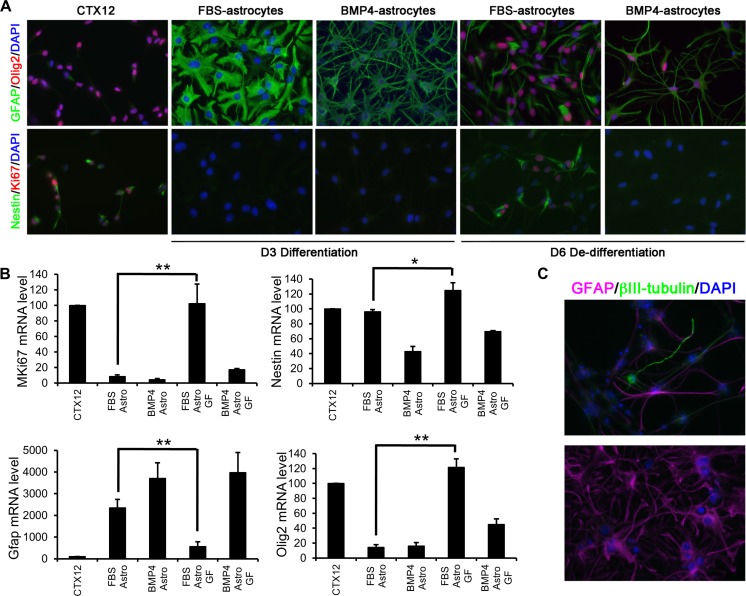
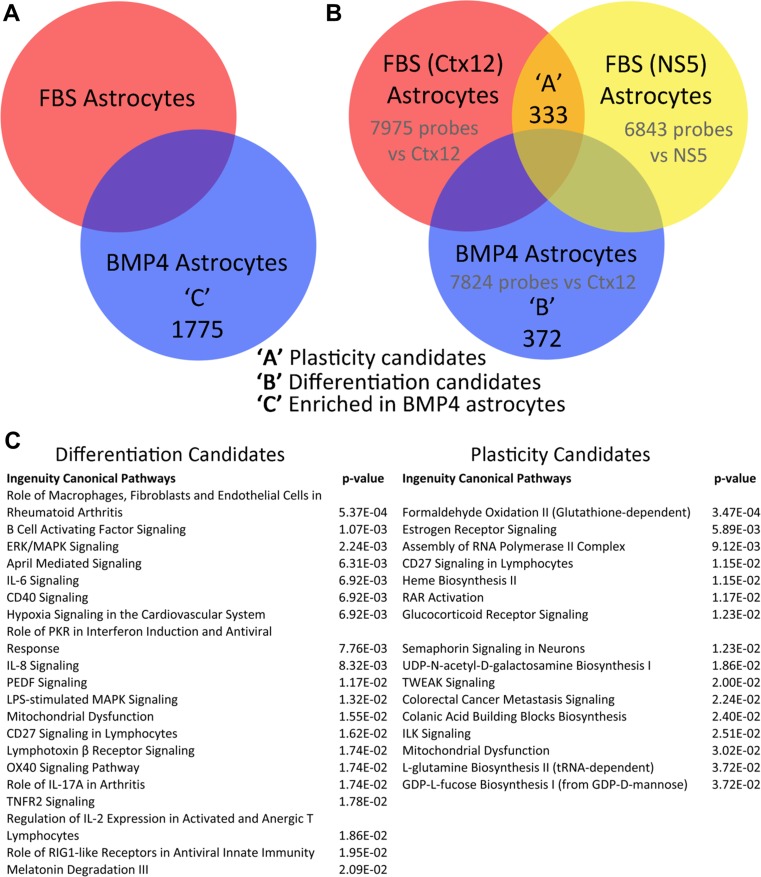
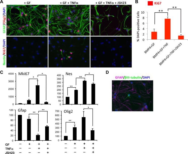
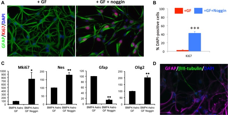
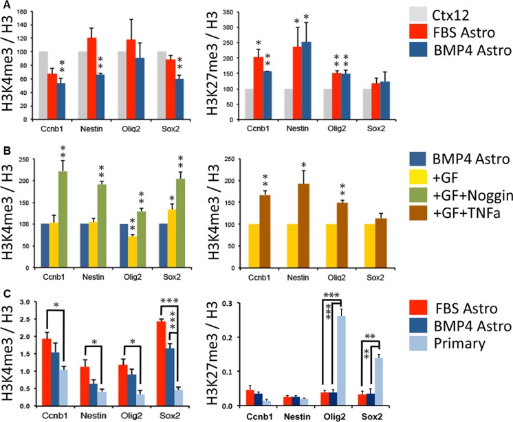
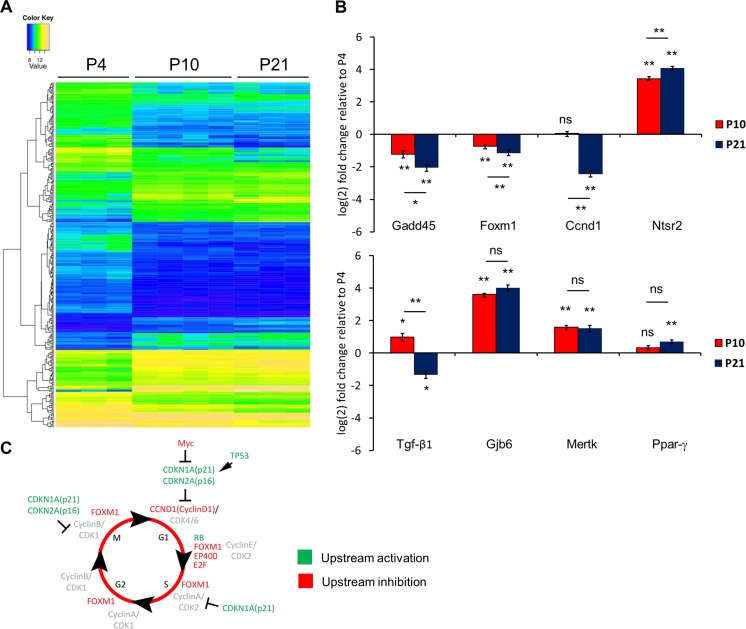
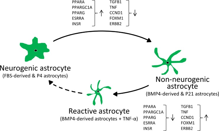
Although the adult brain contains neural stem cells (NSCs) that generate new neurons throughout life, these astrocyte-like populations are restricted to two discrete niches. Despite their terminally differentiated phenotype, adult parenchymal astrocytes can re-acquire NSC-like characteristics following injury, and as such, these 'reactive' astrocytes offer an alternative source of cells for central nervous system (CNS) repair following injury or disease. At present, the mechanisms that regulate the potential of different types of astrocytes are poorly understood. We used in vitro and ex vivo astrocytes to identify candidate pathways important for regulation of astrocyte potential. Using in vitro neural progenitor cell (NPC)-derived astrocytes, we found that exposure of more lineage-restricted astrocytes to either tumor necrosis factor alpha (TNF-α) (via nuclear factor-κB (NFκB)) or the bone morphogenetic protein (BMP) inhibitor, noggin, led to re-acquisition of NPC properties accompanied by transcriptomic and epigenetic changes consistent with a more neurogenic, NPC-like state. Comparative analyses of microarray data from in vitro-derived and ex vivo postnatal parenchymal astrocytes identified several common pathways and upstream regulators associated with inflammation (including transforming growth factor (TGF)-β1 and peroxisome proliferator-activated receptor gamma (PPARγ)) and cell cycle control (including TP53) as candidate regulators of astrocyte phenotype and potential. We propose that inflammatory signalling may control the normal, progressive restriction in potential of differentiating astrocytes as well as under reactive conditions and represent future targets for therapies to harness the latent neurogenic capacity of parenchymal astrocytes.

Keywords: Astrocytes; Epigenetic; Inflammation; NFκB; Neural stem cells; Noggin.


Figures

Fig. 1
Fig. 1
Molecular phenotype of CTX12 cells under proliferative conditions or in the presence of FBS or BMP4. a Immunocytochemistry showing GFAP (green) and Olig2 (red) expression (top panels) and Nestin (green) and Ki67 (red) expression (bottom panels). Nuclei are counterstained with DAPI (blue). CTX12s were differentiated for 3 days (D3) with either FBS or BMP4 followed by 3 days of dedifferentiation (D6). b Comparison of Mki67, Nes, GFAP and Olig2 gene expression in conditions shown in a. c Example of a βIII-tubulin-positive neuron (green) in FBS astrocyte cultures following dedifferentiation and tripotential differentiation (top panel). Bottom panel shows a representative image from BMP4 astrocytes under similar conditions (GFAP in magenta and DAPI in blue). Abbreviations: Astro, astrocyte; GF, growth factors (FGF2/EGF/4-OHT). Error bars in b show SEMs, n = 3
Fig. 2
Fig. 2
Candidate genes and signalling pathways involved in phenotype and potential of specific astrocyte populations. a Venn diagram showing number of genes expressed at a significantly different level between BMP4 and FBS astrocytes. b Venn diagram showing genes whose expressions were significantly changed in FBS- or BMP4-derived astrocytes compared to undifferentiated CTX12 or NS5 cells (FDR < 0.05). c Top canonical pathways enriched in the differentiation candidate gene set ‘B’ and ‘plasticity’ candidate gene set ‘A’ both shown in b
Fig. 3
Fig. 3
A subset of BMP4 astrocytes treated with TNF-α re-enter the cell cycle and re-acquire NPC characteristics. a Immunocytochemistry on BMP4 astrocytes cultured in dedifferentiation conditions for 3 days alone (GF), in the presence of TNF-α (+GF + TNFα) or with TNF-α and JSH23 (+GF + TNFα + JSH23). JSH23 was added 30 min before culturing astrocytes in dedifferentiation conditions in the presence of TNF-α. Top row shows GFAP (green) and Olig2 (red), and bottom row shows Nestin (green) and Ki67 (red). Nuclei were counterstained with DAPI (blue). b Percentage of Ki67-positive cells in cultures shown in a. c Gene expression levels of MKi67, Nestin (Nes), GFAP and Olig2 in BMP4 astrocytes in conditions shown in (a, b). Data are expressed as percentage of expression relative to D3 BMP4 astrocytes. d βIII-tubulin-positive neurons (green) and GFAP-positive astrocytes (magenta) with DAPI-labelled nuclei (blue) in BMP4 astrocyte cultures following dedifferentiation with TNF-α followed by tripotential differentiation. For b and c, n = 3. Scale bars in a and d: 20 μm. P values in b and c: *p < 0.05, **p < 0.01 (Student’s t test), error bars show SEMs
Fig. 4
Fig. 4
Inhibition of BMP signalling leads to re-acquisition of NPC characteristics in BMP4 astrocytes. a Immunocytochemistry comparing Ki67 (red) and GFAP (green) expression in BMP4 astrocytes in dedifferentiation conditions with and without noggin (+GF + noggin and +GF, respectively). Noggin was added simultaneously with GF. Nuclei are counterstained with DAPI (blue). b Percentage of Ki67-positive cells in the conditions shown in a. c Gene expression levels of Mki67, Nes, GFAP and Olig2 in the conditions shown in a. Expression levels are shown as percentage in +GF + noggin conditions relative to +GF conditions (100 %). d βIII-tubulin-positive neurons (green) and GFAP-positive astrocytes (magenta) with DAPI-labelled nuclei (blue) in BMP4 astrocyte cultures following dedifferentiation with noggin followed by tripotential differentiation. For b and c, n = 3. Scale bars in a and d, 20 μm. P values in b and c: *p < 0.05, **p < 0.01, ***p < 0.001 (Student’s t test), error bars show SEMs
Fig. 5
Fig. 5
Epigenetic comparison of phenotypically distinct astrocytes. a H3K4me3 (left) and H3K27me3 (right) enrichment relative to total H3 at specific gene promoters in CTX12 cells (grey), FBS astrocytes (red) and BMP4 astrocytes (blue). Results are expressed as a percentage of levels in CTX12 (100 %). b Left graph shows H3K4me3 enrichment relative to total H3 at specific gene promoters in CTX12 cells (grey) and BMP4 astrocytes after 3 days of dedifferentiation alone (+GF, yellow) or with the addition of noggin (+GF + noggin, green). Results are expressed as a percentage of levels in CTX12 (100 %). The right-hand graph shows a comparison between BMP4 astrocytes in dedifferentiation conditions alone (+GF, yellow) and with TNF-α (+GF + TNFα). c Comparison of H3K4me3 (left) and H3K27me3 (right) enrichment in FBS astrocytes (red), BMP4 astrocytes (dark blue) and primary cortical astrocytes (light blue). For ac, n = 3. *p < 0.05, **p < 0.01, ***p < 0.001 (Student’s t test), and error bars show SEMs
Fig. 6
Fig. 6
Developmental changes in gene expression in postnatal astrocytes. a Clustered heatmap showing relative gene expression levels in P4, P10 and P21 astrocytes from Aldh1l1-EGFP mice. Individual biological replicates are shown as individual columns for P4 (n = 3), P10 (n = 4) and P21 (n = 3). Relative expression levels are shown from low (blue) to high (yellow). b Graphs show Gadd45, Foxm1, Ccnd1, Ntsr2, TGF-β1, Gjb6, Mertk and PPARγ expression levels from FACSed Aldh1l1-EGFP astrocyte populations. Expression levels are shown in P10 (red) and P21 (blue) populations relative to P4 levels (x-axis). Data are the average of three biological replicates. Error bars show SEMs. P values: *p < 0.05, **p < 0.01, ns = not significant (Student’s t test). c Cell cycle diagram. The scheme shows activated (green) or inhibited (red) upstream regulators during in vivo astrocyte maturation as predicted by IPA
Fig. 7
Fig. 7
Candidate upstream regulators in the astrocyte neurogenic/non-neurogenic shift. The scheme illustrates that selected potential regulators that are responsible for the shift of neurogenic astrocytes (FBS-derived and P4 astrocytes) towards non-neurogenic astrocytes (BMP4-derived and P21 astrocytes) are also responsible for the dedifferentiation process under a pro-inflammatory environment (BMP4-derived astrocytes + TNF-α)

References

    1. Zhang Y, Barres BA. Astrocyte heterogeneity: an underappreciated topic in neurobiology. Curr Opin Neurobiol. 2010;20:588–594. doi: 10.1016/j.conb.2010.06.005. - DOI - PubMed
    1. Molofsky AV, Krenick R, Ullian EM, Tsai HH, Deneen B, Richardson WD, Barres BA, Rowitch DH. Astrocytes and disease: a neurodevelopmental perspective. Genes Dev. 2012;26:891–907. doi: 10.1101/gad.188326.112. - DOI - PMC - PubMed
    1. Anderson MA, Ao Y, Sofroniew MV. Heterogeneity of reactive astrocytes. Neurosci Lett. 2014;565:23–29. doi: 10.1016/j.neulet.2013.12.030. - DOI - PMC - PubMed
    1. Doetsch F. A niche for adult neural stem cells. Curr Opin Genet Dev. 2003;13:543–550. doi: 10.1016/j.gde.2003.08.012. - DOI - PubMed
    1. Doetsch F, Caille I, Lim DA, Garcia-Verdugo JM, Alvarez-Buylla A. Subventricular zone astrocytes are neural stem cells in the adult mammalian brain. Cell. 1999;97:703–716. doi: 10.1016/S0092-8674(00)80783-7. - DOI - PubMed

Publication types

MeSH terms

LinkOut - more resources