

High-resolution NMR characterization of low abundance oligomers of amyloid-β without purification
- PMID: 26138908
- PMCID: PMC4490348
- DOI: 10.1038/srep11811
High-resolution NMR characterization of low abundance oligomers of amyloid-β without purification
Abstract
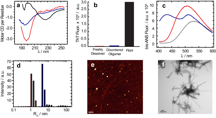
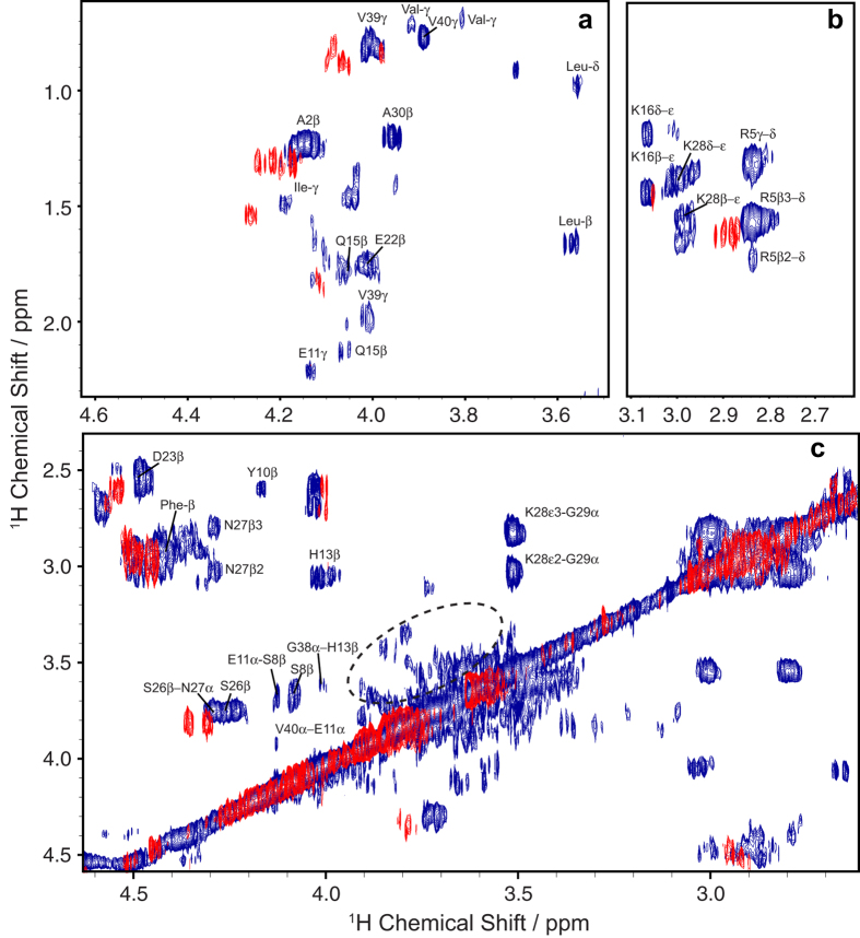
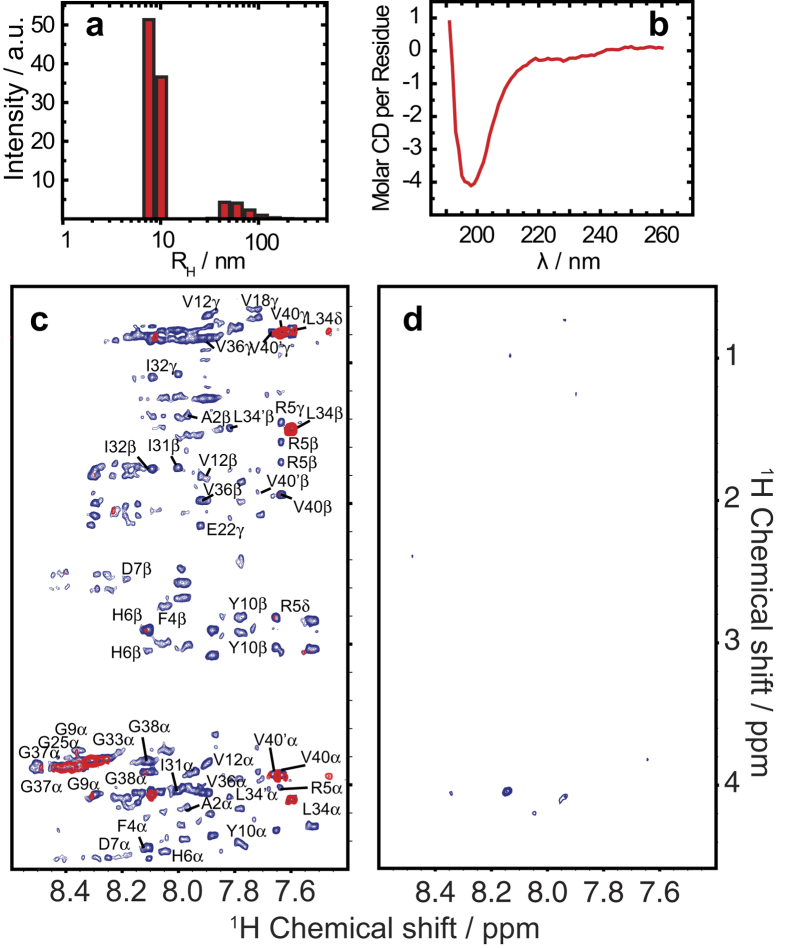
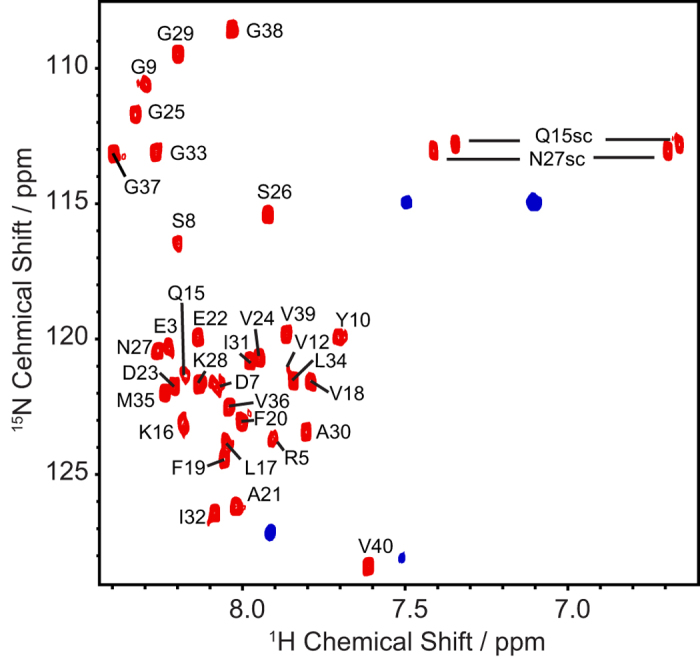
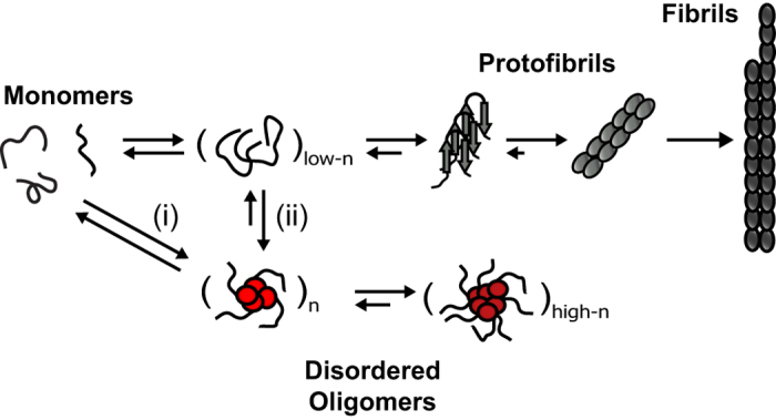
Alzheimer's disease is characterized by the misfolding and self-assembly of the amyloidogenic protein amyloid-β (Aβ). The aggregation of Aβ leads to diverse oligomeric states, each of which may be potential targets for intervention. Obtaining insight into Aβ oligomers at the atomic level has been a major challenge to most techniques. Here, we use magic angle spinning recoupling (1)H-(1)H NMR experiments to overcome many of these limitations. Using (1)H-(1)H dipolar couplings as a NMR spectral filter to remove both high and low molecular weight species, we provide atomic-level characterization of a non-fibrillar aggregation product of the Aβ1-40 peptide using non-frozen samples without isotopic labeling. Importantly, this spectral filter allows the detection of the specific oligomer signal without a separate purification procedure. In comparison to other solid-state NMR techniques, the experiment is extraordinarily selective and sensitive. A resolved 2D spectra could be acquired of a small population of oligomers (6 micrograms, 7% of the total) amongst a much larger population of monomers and fibers (93% of the total). By coupling real-time (1)H-(1)H NMR experiments with other biophysical measurements, we show that a stable, primarily disordered Aβ1-40 oligomer 5-15 nm in diameter can form and coexist in parallel with the well-known cross-β-sheet fibrils.
Figures

Similar articles
-
Antiparallel β-Sheet Structure within the C-Terminal Region of 42-Residue Alzheimer's Amyloid-β Peptides When They Form 150-kDa Oligomers.J Mol Biol. 2015 Jul 3;427(13):2319-28. doi: 10.1016/j.jmb.2015.04.004. Epub 2015 Apr 16. J Mol Biol. 2015. PMID: 25889972 Free PMC article.
-
Supramolecular structural constraints on Alzheimer's beta-amyloid fibrils from electron microscopy and solid-state nuclear magnetic resonance.Biochemistry. 2002 Dec 24;41(51):15436-50. doi: 10.1021/bi0204185. Biochemistry. 2002. PMID: 12484785
-
NMR-based site-resolved profiling of β-amyloid misfolding reveals structural transitions from pathologically relevant spherical oligomer to fibril.J Biol Chem. 2020 Jan 10;295(2):458-467. doi: 10.1074/jbc.RA119.008522. Epub 2019 Nov 26. J Biol Chem. 2020. PMID: 31771980 Free PMC article.
-
High-resolution probing of early events in amyloid-β aggregation related to Alzheimer's disease.Chem Commun (Camb). 2020 Apr 30;56(34):4627-4639. doi: 10.1039/d0cc01551b. Epub 2020 Apr 17. Chem Commun (Camb). 2020. PMID: 32300761 Free PMC article. Review.
-
Inhibition of amyloid-β aggregation in Alzheimer's disease.Curr Pharm Des. 2014;20(8):1223-43. doi: 10.2174/13816128113199990068. Curr Pharm Des. 2014. PMID: 23713775 Review.
Cited by
-
Molecular Mechanism and Kinetics of Amyloid-β42 Aggregate Formation: A Simulation Study.ACS Chem Neurosci. 2019 Nov 20;10(11):4643-4658. doi: 10.1021/acschemneuro.9b00473. Epub 2019 Nov 11. ACS Chem Neurosci. 2019. PMID: 31660732 Free PMC article.
-
Catalytides derived from the Box A region in the ANA/BTG3 protein cleave amyloid-β fragment peptide.Heliyon. 2019 Sep 24;5(9):e02454. doi: 10.1016/j.heliyon.2019.e02454. eCollection 2019 Sep. Heliyon. 2019. PMID: 31687556 Free PMC article.
-
LL-37: Structures, Antimicrobial Activity, and Influence on Amyloid-Related Diseases.Biomolecules. 2024 Mar 8;14(3):320. doi: 10.3390/biom14030320. Biomolecules. 2024. PMID: 38540740 Free PMC article. Review.
-
Exploiting heterogeneous time scale of dynamics to enhance 2D HETCOR solid-state NMR sensitivity.J Magn Reson. 2019 Dec;309:106615. doi: 10.1016/j.jmr.2019.106615. Epub 2019 Oct 14. J Magn Reson. 2019. PMID: 31669793 Free PMC article.
-
A High Affinity Red Fluorescence and Colorimetric Probe for Amyloid β Aggregates.Sci Rep. 2016 Apr 1;6:23668. doi: 10.1038/srep23668. Sci Rep. 2016. PMID: 27032526 Free PMC article.
References
-
- Alzheimer’s disease facts and figures. Alzheimer’s Dement. 11, 332–384 (2015). - PubMed
-
- Sakono M. & Zako T. Amyloid oligomers: formation and toxicity of Abeta oligomers. FEBS J. 277, 1348–58 (2010). - PubMed
-
- Benilova I., Karran E. & De Strooper B. The toxic Aβ oligomer and Alzheimer’s disease: an emperor in need of clothes. Nat. Neurosci. 15, 349–57 (2012). - PubMed
-
- Selkoe D. J. The molecular pathology of Alzheimer’s disease. Neuron 6, 487–498 (1991). - PubMed
-
- Lin H., Bhatia R. & Lal R. Amyloid beta protein forms ion channels: implications for Alzheimer’s disease pathophysiology. FASEB J 15, 2433–2444 (2001). - PubMed
Publication types
MeSH terms
Substances
LinkOut - more resources
Full Text Sources
Other Literature Sources
Medical
