Review
. 2015 Jul 1;87(1):63-75.
doi: 10.1016/j.neuron.2015.05.043.

Biochemical Computation for Spine Structural Plasticity

Affiliations
Review

Biochemical Computation for Spine Structural Plasticity

Jun Nishiyama et al. Neuron. .

Abstract

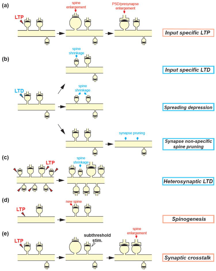
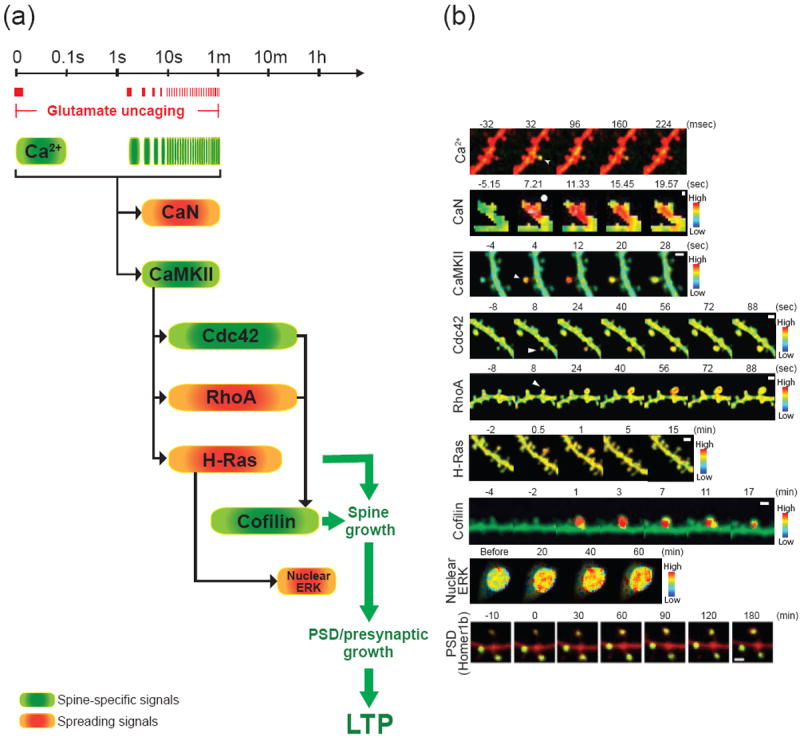
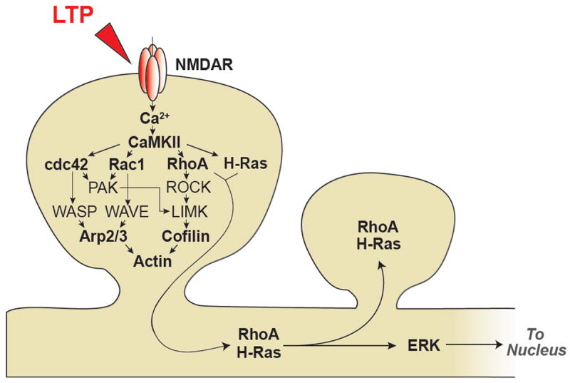
The structural plasticity of dendritic spines is considered to be essential for various forms of synaptic plasticity, learning, and memory. The process is mediated by a complex signaling network consisting of numerous species of molecules. Furthermore, the spatiotemporal dynamics of the biochemical signaling are regulated in a complicated manner because of geometrical restrictions from the unique morphology of the dendritic branches and spines. Recent advances in optical techniques have enabled the exploration of the spatiotemporal aspects of the signal regulations in spines and dendrites and have provided many insights into the principle of the biochemical computation that underlies spine structural plasticity.


Figures

Fig.1
Fig.1
Spine structural plasticity (a) Structural plasticity during LTP: Repetitive 2-photon uncaging of MINI-glutamate (0.5-2Hz for 1min) at a single spine under low Mg2+ (nominally zero) condition or paired with postsynaptic depolarization induces a rapid enlargement of the spine head in a few minutes. The volume of the enlarged spine gradually decreases over ~5 min to a plateau and sustains more than an hour (Lee et al., 2009; Matsuzaki et al., 2004). The PSD and presynapse increase with a delay of 0.5-3 hours (Bosch et al., 2014; Meyer et al., 2014). Note that the enlargement of the spine volume is restricted to the stimulated spine (input specific LTP). (b) Structural plasticity during LTD: Different protocols for LTD induction have been reported to result in the distinct structural plasticity. Low frequency glutamate uncaging (90 pulses at 0.1Hz) in low extracellular Ca2+ (0.3mM) and Mg2+ (nominally zero) concentration or paired with postsynaptic depolarization induces a spine shrinkage restricted to the stimulated spine (input specific LTD) (Oh et al., 2013) (upper). B-AP paired with subsequent 2-photon glutamate uncaging pulses (~10 ms) at a single spine (80 pulses at 1Hz) shortly after (< 50 ms) GABA uncaging at the adjacent dendritic shaft induces the reduction in the volume of the stimulated spine as well as neighboring non-stimulated spines (spreading depression) (Hayama et al., 2013) (middle). The optogenetic stimulation of presynaptic CA3 pyramidal neurons expressing channelrodopsin-2 (1Hz for 900 light pulses) induces functional LTD but not spine shrinkage in postsynaptic CA1 neurons. However, a few days later, the stimulated spine and many neighboring synapses are removed (synapse-nonspecific spine pruning) (Wiegert and Oertner, 2013) (lower). (c) Heterosynaptic LTD: LTP stimulation at multiple spines on a single dendritic segment by glutamate uncaging induces shrinkage of nearby unstimulated spines (Oh et al., 2015). (d) Spinogenesis induced by glutamate uncaging: 2-photon glutamate uncaging (40 pulses at 2Hz) at dendritic shafts triggers rapid de novo spinogenesis in young neurons (Kwon and Sabatini, 2011). (e) Synaptic crosstalk associated with structural plasticity: Repetitive glutamate uncaging (30 pulses at 0.5Hz, 4ms pulse duration) is applied to a single spine to induce LTP. A subthreshold stimulus (30 pulses at 0.5Hz, 1ms pulse duration), which by itself does not trigger LTP, is then applied to a nearby spine. This induces a sustained structural and functional LTP in the weakly stimulated spine (Harvey and Svoboda, 2007; Harvey et al., 2008).
Fig.2
Fig.2
The spatiotemporal dynamics of signaling activities during structural LTP (a) The timescale of signaling activities during structural LTP induced by 2-photon glutamate uncaging (0.5 - 20 Hz). Spine specific signals and spreading signals are indicated in green and orange, respectively. The timings of glutamate uncaging of a typical LTP induction protocol (0.5 Hz) are shown in red bars. (b) Ca2+ elevation, activities of CaN, CaMKII, cdc42, RhoA, H-Ras, cofilin, nuclear ERK and the accumulation of Homer1b during structural LTP induced at a single spine or 7 spines (ERK). The arrows and circles show the spines stimulated with glutamate uncaging. Scale bars (white) are 10 μm for ERK and 1 μm for others. The images are adopted and modified from (Zhai et al., 2013) for Ca2+ and ERK, (Fujii et al., 2013) for CaN, (Lee et al., 2009) for CaMKII, (Murakoshi et al., 2011) for RhoA and cdc42, (Harvey et al., 2008) for H-Ras and (Bosch et al., 2014) for cofilin and Homer1b with permission. The Ca2+ elevation is visualized with a Ca2+ indicator Fluo-4FF (green) and Alexa-594 (red). Note that Ca2+ elevation in response to the first uncaging pulse during LTP induction protocol (1 Hz, 60 pulses) is displayed. CaMKII, cdc42, RhoA, H-Ras, cofilin and nuclear ERK activities are imaged with 2pFLIM combined with FRET sensors. CaN activities are visualized with dual FRET with optical manipulation (dFOMA). The accumulation of Homer1b in spines is visualized with GFP tagged-Homer1b and RFP (cell fill). Note that the Ca2+ elevation and the activations of CaMKII, cdc42, and cofilin are restricted to the stimulated spines, whereas the activation of CaN, RhoA and H-Ras spread into the dendritic shafts and the nearby spines.
Fig.3
Fig.3
Schematic diagram of signaling pathways associated with structural LTP

References

    1. Aakalu G, Smith WB, Nguyen N, Jiang C, Schuman EM. Dynamic visualization of local protein synthesis in hippocampal neurons. Neuron. 2001;30:489–502. - PubMed
    1. Abraham WC. Metaplasticity: tuning synapses and networks for plasticity. Nature reviews Neuroscience. 2008;9:387. - PubMed
    1. Abraham WC, Christie BR, Logan B, Lawlor P, Dragunow M. Immediate early gene expression associated with the persistence of heterosynaptic long-term depression in the hippocampus. Proceedings of the National Academy of Sciences of the United States of America. 1994;91:10049–10053. - PMC - PubMed
    1. Adams JP, Dudek SM. Late-phase long-term potentiation: getting to the nucleus. Nature reviews Neuroscience. 2005;6:737–743. - PubMed
    1. Alberini CM. Transcription factors in long-term memory and synaptic plasticity. Physiological reviews. 2009;89:121–145. - PMC - PubMed

Publication types

LinkOut - more resources