. 2015 Jul 1;87(1):220-30.
doi: 10.1016/j.neuron.2015.06.016.

Rapid Encoding of New Memories by Individual Neurons in the Human Brain

Affiliations

Rapid Encoding of New Memories by Individual Neurons in the Human Brain

Matias J Ison et al. Neuron. .

Abstract

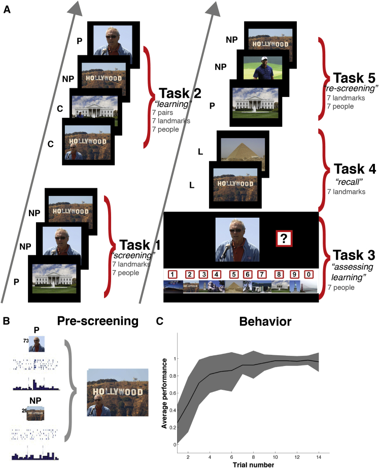
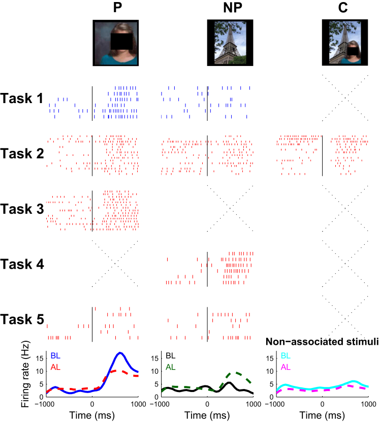
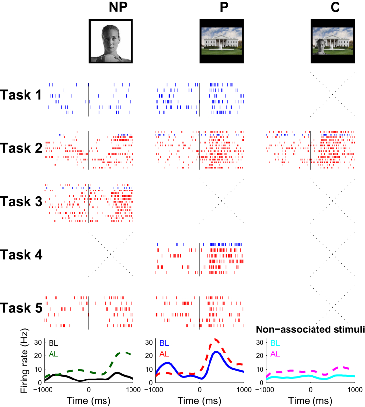
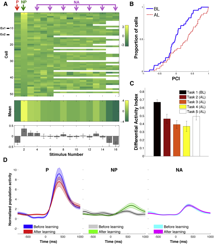
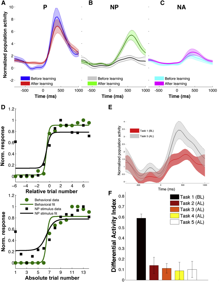
The creation of memories about real-life episodes requires rapid neuronal changes that may appear after a single occurrence of an event. How is such demand met by neurons in the medial temporal lobe (MTL), which plays a fundamental role in episodic memory formation? We recorded the activity of MTL neurons in neurosurgical patients while they learned new associations. Pairs of unrelated pictures, one of a person and another of a place, were used to construct a meaningful association modeling the episodic memory of meeting a person in a particular place. We found that a large proportion of responsive MTL neurons expanded their selectivity to encode these specific associations within a few trials: cells initially responsive to one picture started firing to the associated one but not to others. Our results provide a plausible neural substrate for the inception of associations, which are crucial for the formation of episodic memories.


Figures

Figure 1
Figure 1
Experimental Design and Behavioral Results (A) Structure of the association task. P, preferred stimulus; NP, non-preferred stimulus; C, composite stimulus; L, landmarks. (B) Selection of stimuli. In this example, in a previous recording session (performed prior to the tasks in A to determine pictures eliciting responses in the neurons), we identified one single unit that responded to a picture of the American actor Clint Eastwood (P) and did not change its firing rate in response to the picture of the Hollywood sign (NP). The preferred (P) and non-preferred (NP) stimuli for each neuron were used to create contextual pictures as the one shown (median 7 pairs, 3–8 pairs per session). (C) Grand average learning curve (mean ± SD) for all pairs in 25 sessions performed by 14 patients. Trial number refers to trials during Task 3, where learning was assessed. Note the high variability across sessions.
Figure 2
Figure 2
Exemplary Response in the Hippocampus A unit in the left hippocampus of participant 14 was activated with a response of 13.1 spikes/s when the image of the patient’s family was presented (preferred stimulus, black squares have been added for privacy reasons). The same cell was not responsive (response: 3.3 spikes/s) to the image of the Eiffel tower before learning (Task 1). For each task the corresponding raster plots (ordered from top to bottom) of each picture are given. Blue rasters represent pre-learning (Task 1) or incorrect trials. Red rasters represent correct or post-learning (Task 5) trials. The spike density function for trials before (BL) and after (AL) learning in response to the non-preferred (left), preferred (middle), and to the mean of the non-associated stimuli (average over 7 pictures) are shown at the bottom panels. Crosses indicate that the stimulus was not shown during a given task. After single-trial learning (Tasks 2, 3, and 4), the unit fired strongly to the picture of the patient’s family (mean: 10.8 spikes/s, left), to the composite picture (7.8 spikes/s, right) and to the picture of the Eiffel tower (7.6 spikes/s). There was a 230% increase in firing to the non-preferred stimulus. The response to the non-associated stimuli slightly decreased from 5.3 spikes/s before learning to 3.6 spikes/s after learning.
Figure 3
Figure 3
Exemplary Response in the Parahippocampal Cortex Conventions are the same as in Figure 2. A multi-unit in the parahippocampal cortex of participant 3 fired at a rate of 17.8 spikes/s (SD = 7.2) to the picture of the White House (preferred stimulus) from a baseline of 4.4 spikes/s (SD = 4.0). This cell only fired at a rate of 5.0 spikes/s (SD = 3.6) to the picture of the American volleyball player Kerri Walsh before learning (Task 1). After learning (trial 1 in Task 2), the cell selectively increased (by 246%) its response to the pair associate (mean response: 13.8 spikes/s, SD = 9.1, p < 0.05).
Figure 4
Figure 4
Population: Visually Responsive Units (A) Response changes for all visually responsive units. Each row represents one cell and each column represents one stimulus. The rows were sorted by the strength of the change in the NP stimulus and the columns were unsorted. Blank squares represent stimuli that were not shown during the corresponding session. The mean values across all cells are shown in the middle panel (in colors) and in the bottom panel including SEMs. Ex1, Ex2 correspond to the exemplary units shown in Figures 2 and 3. (B) Cumulative frequency histograms of the correlation coefficient (defined as in Higuchi and Miyashita, 1996) for units before learning (BL) and after learning (AL). Correlation coefficients were significantly higher after learning than before learning (p = 0.007, Kolmogorov-Smirnov test). (C) Average differential activity index DAI = (Pr − NPr / Pr + NPr) for all tasks. Lower values of DAI denote more similar responses. Responses to the preferred and non-preferred stimuli become more similar after learning for all tasks (p < 0.001, average decrease by a factor of 1.6, range: 1.4–1.8). (D) Average normalized spike density function (SDF) for 51 visually responsive units to the P, NP, and NA before and after learning. There was a significant increase in the response strength to the NP stimuli after learning (p < 0.05, Wilcoxon rank-sum test).
Figure 5
Figure 5
Population: Pair-Coding Units Average normalized spike density function (SDF) for 21 units that selectively changed their response after learning. The shaded areas represent SEM. (A) Normalized SDF to the preferred stimulus for before learning (BL) and after learning (AL). There was no significant difference between conditions in the response period. (B) Normalized SDF to the non-preferred stimulus for BL and AL. After learning, units responded significantly more strongly to the non-preferred stimulus (p < 0.01). (C) Normalized SDF to the non-associated stimuli. (D) Average normalized neural activity (black squares) and behavioral responses (green circles) to the non-preferred stimulus as a function of trial number. In the top panel, data were aligned to the learning time (relative trial number 0). In the bottom panel, trials were sorted according to their presentation order, with the first 6 trials always denoting trial 1 and trial 7 corresponding to the start of Task 2. Continuous lines correspond to psychometric fits using a binomial function. Note that the neural activity follows the sudden increase in behavioral learning when data are aligned relative to learning time. (E) Normalized SDF to the non-preferred stimulus for Task 1 and Task 5. Responses during Task 5 were significantly higher than during Task 1 (p = 0.001, Wilcoxon rank-sum test). (F) Average differential activity index DAI for all tasks. Responses to the preferred and non-preferred stimuli become more similar after learning for all tasks (p < 10−6, average decrease by a factor of 5.5, range: 4.2–6.7).

References

    1. Akaike H. A new look at the statistical model identification. IEEE Trans. Auto. Contr. 1974;19:716–723.
    1. Aminoff E., Gronau N., Bar M. The parahippocampal cortex mediates spatial and nonspatial associations. Cereb. Cortex. 2007;17:1493–1503. - PubMed
    1. Buffalo E.A., Bellgowan P.S., Martin A. Distinct roles for medial temporal lobe structures in memory for objects and their locations. Learn. Mem. 2006;13:638–643. - PMC - PubMed
    1. Bunsey M., Eichenbaum H. Conservation of hippocampal memory function in rats and humans. Nature. 1996;379:255–257. - PubMed
    1. Davachi L. Item, context and relational episodic encoding in humans. Curr. Opin. Neurobiol. 2006;16:693–700. - PubMed

Publication types

MeSH terms