. 2015 Jul 3:5:11645.
doi: 10.1038/srep11645.

Structural colour in Chondrus crispus

Affiliations

Structural colour in Chondrus crispus

Chris J Chandler et al. Sci Rep. .

Abstract

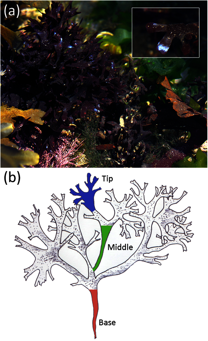
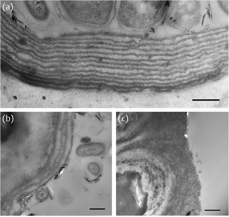
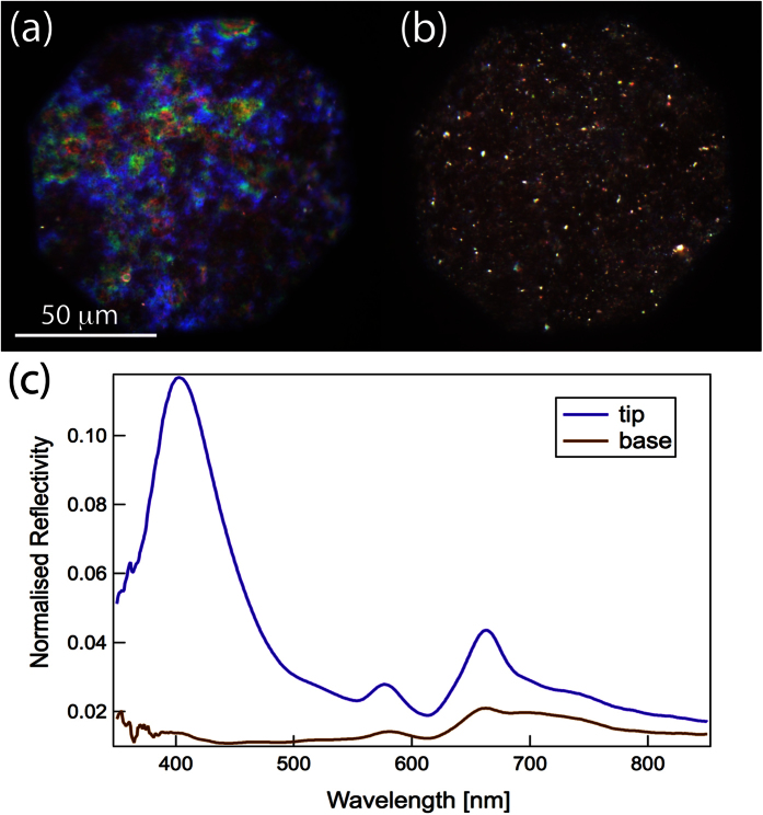
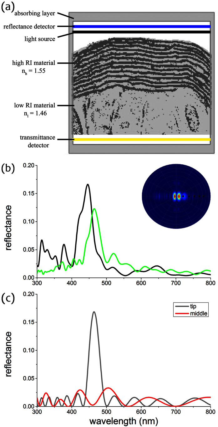
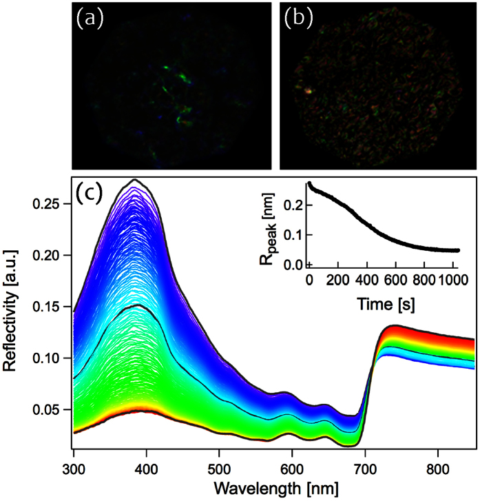
The marine world is incredibly rich in brilliant and intense colours. Photonic structures are found in many different species and provide extremely complex optical responses that cannot be achieved solely by pigments. In this study we examine the cuticular structure of the red alga Chondrus crispus (Irish Moss) using anatomical and optical approaches. We experimentally measure the optical response of the multilayer structure in the cuticle. Using finite-difference time-domain modelling, we demonstrate conclusively for the first time that the dimensions and organisation of lamellae are responsible for the blue structural colouration on the surface of the fronds. Comparison of material along the apical-basal axis of the frond demonstrates that structural colour is confined to the tips of the thalli and show definitively that a lack of structural colour elsewhere corresponds with a reduction in the number of lamellae and the regularity of their ordering. Moreover, by studying the optical response for different hydration conditions, we demonstrate that the cuticular structure is highly porous and that the presence of water plays a critical role in its ability to act as a structural light reflector.


Conflict of interest statement

The authors declare no competing financial interests.

Figures

Figure 1
Figure 1
(a) Habitat view of Irish moss, Chondrus crispus. The intense blue colour at the tip of the thalli is clearly visible and strongly contrasts with the purple-red colour of the frond. (b) Illustration of a typical Chondrus crispus sample highlighting the position of the different sections of the thalli investigated in this study; base (red), middle (green) and tip (blue). All photographs in this figure were taken by Chris J. Chandler.
Figure 2
Figure 2
TEM micrographs showing the cuticle of a Chondrus crispus frond at sections from (a) tip, (b) middle and (c) base. Scale bars represent 500 nm (a,b), 2 μm (c); optical zoom, (a) 7000x, (b) 4000x, (c) 6000x.
Figure 3
Figure 3
(a,b) Micrographs of Chondrus crispus at the (a) tip and (b) base of the thalli. (c) Reflectance spectra collected in confocal configuration under the microscope in the same area as images (a) and (b).
Figure 4
Figure 4. FDTD simulations.
(a) Binarized version of the TEM image in Fig. 2a in the modelling volume assigned in the FDTD simulations. The light cellulosic material was assigned a refractive index of 1.46, whereas the dark-stained material was assigned a refractive index of 1.55. The boundary box was filled with water, RI = 1.33 (see also Materials & Methods). The outlined area indicates the computational domain with absorbing boundaries, the black bar the position of the light source and the blue and yellow bars the reflectance and transmittance detector, respectively. (b) Simulated reflectance spectra for non-polarized light at normal incidence from FDTD simulations of two different TEM images (green line for structure of Figs 2a and 4a; black line for structure of another TEM image). Inset: Simulated light scattering pattern shows a strong directionality of reflected light. (c) Reflectance spectra of an idealised classical multilayer model for the structure in the tip (gray line) and the middle of the frond (red line).
Figure 5
Figure 5. Loss of structural colour during the evaporation of water.
(a) Wet tissue producing intense structural colouration, (b) dry tissue and loss of colour intensity. (c) Reflectance spectra of Chondrus crispus showing a reduction in peak reflectivity over time. Spectra were collected over 750 seconds at regular intervals of five seconds during the evaporation of the water. Different colour spectra represent different time intervals; wet tissue corresponds with blue spectra, dry corresponds with red spectra. Peak reflectance, 385 nm; measurements were performed under 0° incidence. Inset: the loss of colour intensity at the peak reflectance over time.

References

    1. Chapman R. F. Visual signals: color and light production in The insects: structure and function (eds Vukusic P. & Chittka L.) 793–823 (Cambridge University Press, 1998).
    1. Parker A. R. 515 million years of structural colour. J. Opt. A: Pure Appl. Opt. 2, R15–R28, http://iopscience.iop.org/1464-4258/2/6/201 (2000).
    1. Seago A. E., Brady P., Vigneron J.-P. & Schultz T. D. Gold bugs and beyond: a review of iridescence and structural colour mechanisms in beetles (Coleoptera). J. R. Soc. Interface 6, S165–S184, 10.1098/rsif.2008.0354.focus (2009). - DOI - PMC - PubMed
    1. Vukusic P. & Sambles J. R. Photonic structures in biology. Nature 424, 852–855, 10.1038/nature01941 (2003). - DOI - PubMed
    1. Kinoshita S. & Yoshioka S. Structural colors in nature: the role of regularity and irregularity in the structure. ChemPhysChem 6, 1442–1459, 10.1002/cphc.200500007 (2005). - DOI - PubMed

Publication types

LinkOut - more resources