. 2015 Aug 28;290(35):21352-64.
doi: 10.1074/jbc.M115.667337. Epub 2015 Jul 2.

Structural Basis for the Interconversion of Maltodextrins by MalQ, the Amylomaltase of Escherichia coli

Affiliations

Structural Basis for the Interconversion of Maltodextrins by MalQ, the Amylomaltase of Escherichia coli

Simon C Weiss et al. J Biol Chem. .

Abstract

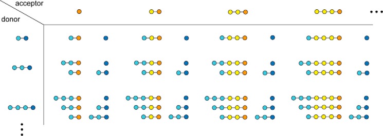
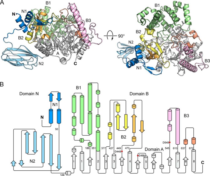
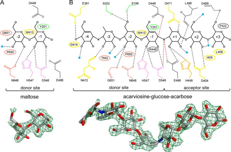
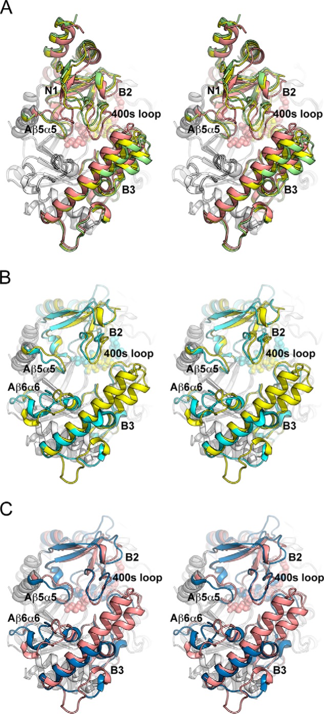
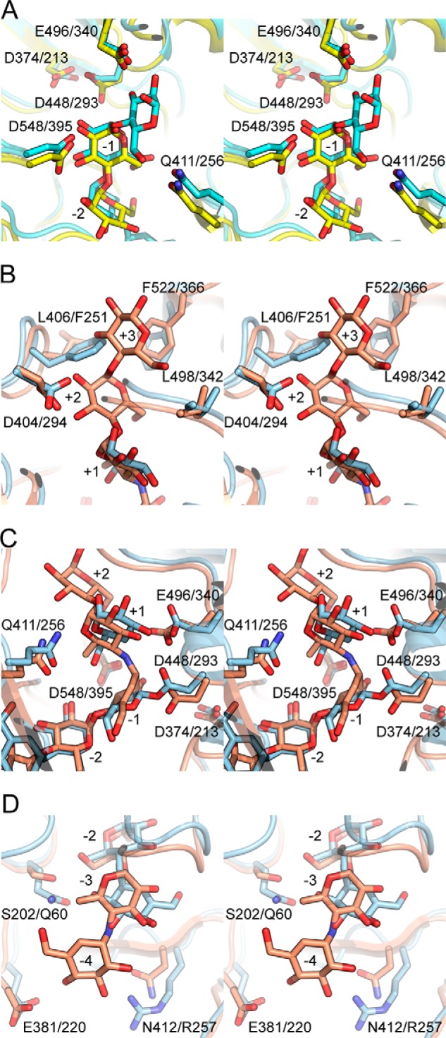
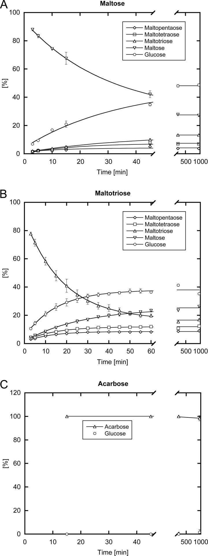
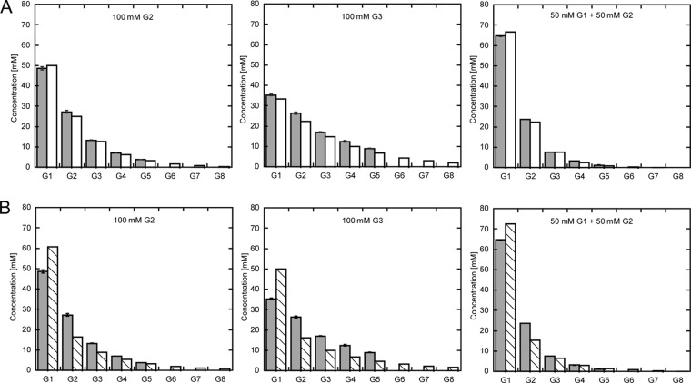
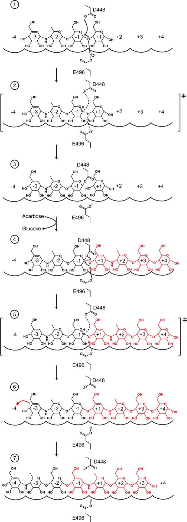
Amylomaltase MalQ is essential for the metabolism of maltose and maltodextrins in Escherichia coli. It catalyzes transglycosylation/disproportionation reactions in which glycosyl or dextrinyl units are transferred among linear maltodextrins of various lengths. To elucidate the molecular basis of transglycosylation by MalQ, we have determined three crystal structures of this enzyme, i.e. the apo-form, its complex with maltose, and an inhibitor complex with the transition state analog acarviosine-glucose-acarbose, at resolutions down to 2.1 Å. MalQ represents the first example of a mesophilic bacterial amylomaltase with known structure and exhibits an N-terminal extension of about 140 residues, in contrast with previously described thermophilic enzymes. This moiety seems unique to amylomaltases from Enterobacteriaceae and folds into two distinct subdomains that associate with different parts of the catalytic core. Intriguingly, the three MalQ crystal structures appear to correspond to distinct states of this enzyme, revealing considerable conformational changes during the catalytic cycle. In particular, the inhibitor complex highlights the requirement of both a 3-OH group and a 4-OH group (or α1-4-glycosidic bond) at the acceptor subsite +1 for the catalytically competent orientation of the acid/base catalyst Glu-496. Using an HPLC-based MalQ enzyme assay, we could demonstrate that the equilibrium concentration of maltodextrin products depends on the length of the initial substrate; with increasing numbers of glycosidic bonds, less glucose is formed. Thus, both structural and enzymatic data are consistent with the extremely low hydrolysis rates observed for amylomaltases and underline the importance of MalQ for the metabolism of maltodextrins in E. coli.

Keywords: Escherichia coli (E. coli); acarbose; carbohydrate metabolism; enzymatically derived inhibitor; enzyme mechanism; maltose/maltodextrin metabolism; protein crystallization; x-ray crystallography.


Figures

FIGURE 1.
FIGURE 1.
Schematic representation of possible MalQ reactions, starting with maltose as the first possible donor for a sugar unit. For clarity, only glucose and the shorter saccharides maltose, maltotriose, and maltotetraose are considered here. Donor and acceptor molecules are depicted in light blue and yellow, respectively, with their reducing ends highlighted in blue and orange. Notably, the primary reaction products are substrates for subsequent reaction cycles (cf. supplemental material).
FIGURE 2.
FIGURE 2.
Structural overview of MalQ. A, schematic representation of apo-MalQ. The TIM barrel domain A, subdomains N1, N2, and B1 to B3 are colored gray, blue, light blue, and green, yellow/orange, pink/salmon, respectively. B, corresponding topology diagram colored as above. Residue numbers indicate the domain boundaries. Red dots mark catalytically important residues.
FIGURE 3.
FIGURE 3.
Ligand interactions in the MalQ·maltose (A) and MalQ·AGA complexes (B). Schematic representations of the ligands (black) and all interacting residues (colored as in Fig. 2). MalQ residues that form hydrogen bonds or van der Waals contacts with the respective ligand are indicated as sticks and ellipses, respectively. Water molecules that interact with both the ligand and the protein are depicted as small blue spheres. Numbers inside the sugar rings label the corresponding subsites. Contacts between the sugar substrate and the nucleophilic side chain of Asp-448 (2.6 Å), the acid/base catalyst Glu-496 (2.9 Å), and the putative transition state stabilizer Asp-548 (3.0 Å) are highlighted as red dashed lines. In the MalQ·maltose structure these residues are too far apart (Asp-448, 4.2 Å; Glu-496, 6.0 Å) for contact formation with the substrate. Illustration of the omit FOFC electron density map (green mesh; contoured at 2.0 and 3.0 σ for maltose and AGA, respectively) with superimposed ligand (stick model) is depicted underneath.
FIGURE 4.
FIGURE 4.
Structural comparison (stereo view) of MalQ with amylomaltase from Thermus sp. A, superposition of the MalQ structures in schematic representation as follows: apo-MalQ (green), MalQ·maltose (yellow), and MalQ·AGA (salmon). Subdomain N2 was omitted for clarity. B, superposition of MalQ·maltose (yellow) and amylomaltase from T. aquaticus (PDB code 1ESW, cyan) (40). Subdomains N1 and N2 of MalQ are not depicted, due to their absence in the Thermus structures. C, similar superposition of MalQ·AGA (salmon) and the amylomaltase of T. thermophilus (PDB code 2OWW, blue) (19). For superposition, the 36 Cα positions of the eight β-strands within the central TIM barrel were used, corresponding to residues 143–148, 181–184, 370–375, 444–447, 492–495, 516–519, 540–543, and 632–635 of MalQ. Ligands are shown as spheres in the same color as the schematics.
FIGURE 5.
FIGURE 5.
Comparison of MalQ·ligand complexes (stereo view) with those of amylomaltases from Thermus sp. A, MalQ·maltose (yellow) versus TaAM·acarbose (cyan, 1ESW), subsites −2 to −1. B–D, comparison of the subsites of MalQ·AGA (salmon) versus TtAM·AG·4G (blue, 2OWW), subsites +3 to +1 (B), subsites +2 to −2 (C), and subsites −2 to −4 (D). Ligands and relevant residues are shown as sticks in the same color.
FIGURE 6.
FIGURE 6.
MalQ enzymatic assay. Evolution of maltodextrin product concentrations including glucose, maltose, maltotriose, maltotetraose, and maltopentaose starting from the substrates maltose (A) and maltotriose (B) are plotted over time. C, illustrates the very slow conversion of acarbose by MalQ. The product mixtures were derivatized with dansylhydrazine, separated by HPLC and their relative peak areas were quantified.
FIGURE 7.
FIGURE 7.
Equilibrium concentrations for different maltodextrin substrates. Experimentally determined maltodextrin equilibrium concentrations comprising G1 (glucose) to G8 (maltooctaose) for the substrates 100 mm maltose, 100 mm maltotriose, and a mixture of 50 mm maltose + 50 mm glucose are shown as gray bars. A, comparison of experimental values with values according to the empirical formula (cf. “Results”; white bars). B, predicted statistical product distribution (cf. “Experimental Procedures”; white striped bars) in comparison with the experimental values (same as in A).
FIGURE 8.
FIGURE 8.
Mechanism of MalQ inhibition by acarbose. The two acarbose molecules that are converted to AGA by MalQ are colored black and red. MalQ subsites are indicated by arches and numbered −4 to +4. The catalytic residues Asp-448 and Glu-496 are shown as sticks. The reaction proceeds in seven stages (see text), each shown in one panel, which can be deduced from distinct crystal structures. Panel 1, MalQ·maltose and TaAM·acarbose (PDB code 1ESW); panel 2, MalQ·AGA; panels 3 and 4, TtAM·AG and TtAM·AG·4G (PDB codes 2OWC and 2OWW); panel 5, MalQ·AGA; panel 6, MalQ·maltose and TaAM·acarbose (PDB code 1ESW); and panel 7, MalQ·AGA.

References

    1. Boos W., Shuman H. (1998) Maltose/maltodextrin system of Escherichia coli: transport, metabolism, and regulation. Microbiol. Mol. Biol. Rev. 62, 204–229 - PMC - PubMed
    1. Freundlieb S., Ehmann U., Boos W. (1988) Facilitated diffusion of p-nitrophenyl-α-d-maltohexaoside through the outer membrane of Escherichia coli. Characterization of LamB as a specific and saturable channel for maltooligosaccharides. J. Biol. Chem. 263, 314–320 - PubMed
    1. Xia T., Han Q., Costanzo W. V., Zhu Y., Urbauer J. L., Eiteman M. A. (2015) Accumulation of d-glucose from pentoses by Escherichia coli. Appl. Environ. Microbiol. 81, 3387–3394 - PMC - PubMed
    1. Becker S., Palm D., Schinzel R. (1994) Dissecting differential binding in the forward and reverse reaction of Escherichia coli maltodextrin phosphorylase using 2-deoxyglucosyl substrates. J. Biol. Chem. 269, 2485–2490 - PubMed
    1. O'Reilly M., Watson K. A., Schinzel R., Palm D., Johnson L. N. (1997) Oligosaccharide substrate binding in Escherichia coli maltodextrin phosphorylase. Nat. Struct. Biol. 4, 405–412 - PubMed

Publication types

MeSH terms

LinkOut - more resources