. 2015 Aug 28;290(35):21409-20.
doi: 10.1074/jbc.M114.634162. Epub 2015 Jul 2.

Nicotinic Acetylcholine Receptors Sensitize a MAPK-linked Toxicity Pathway on Prolonged Exposure to β-Amyloid

Affiliations

Nicotinic Acetylcholine Receptors Sensitize a MAPK-linked Toxicity Pathway on Prolonged Exposure to β-Amyloid

Komal Arora et al. J Biol Chem. .

Abstract

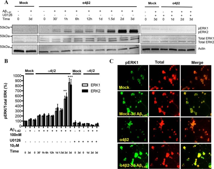
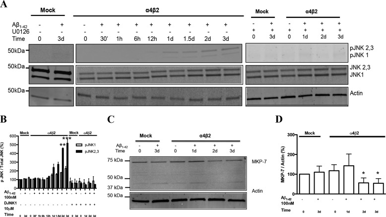
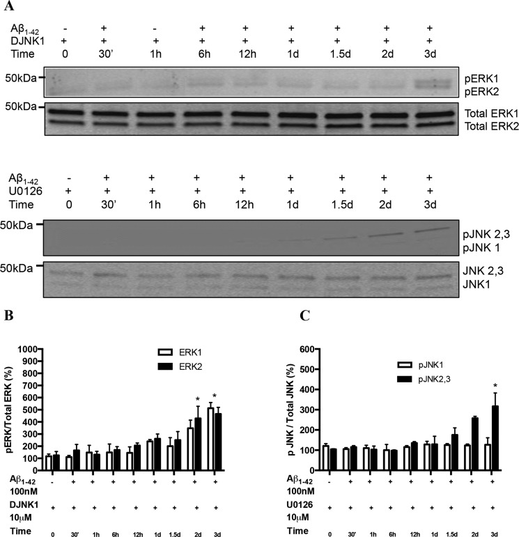
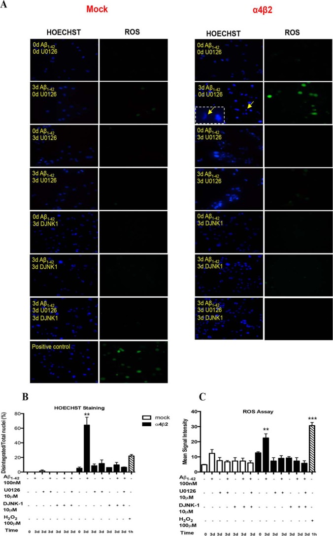
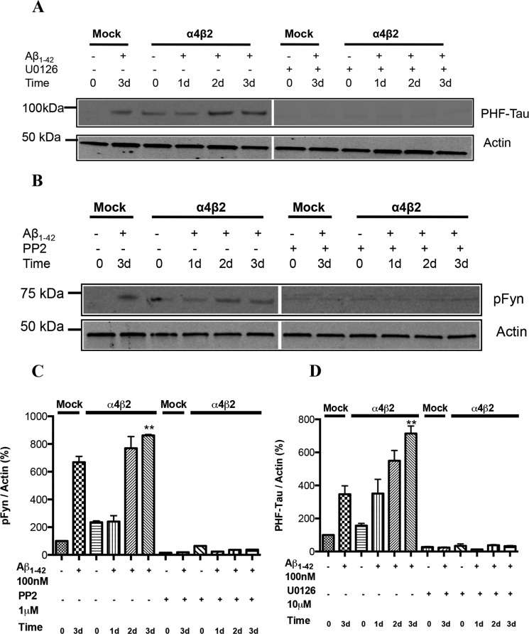
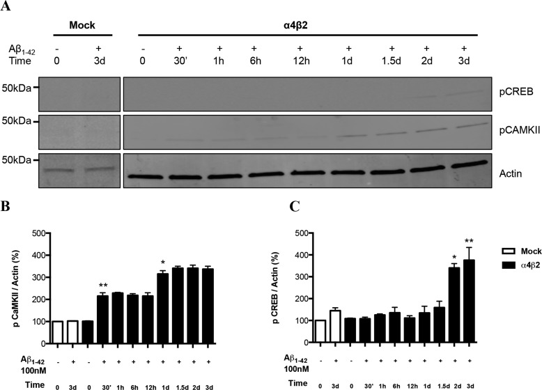
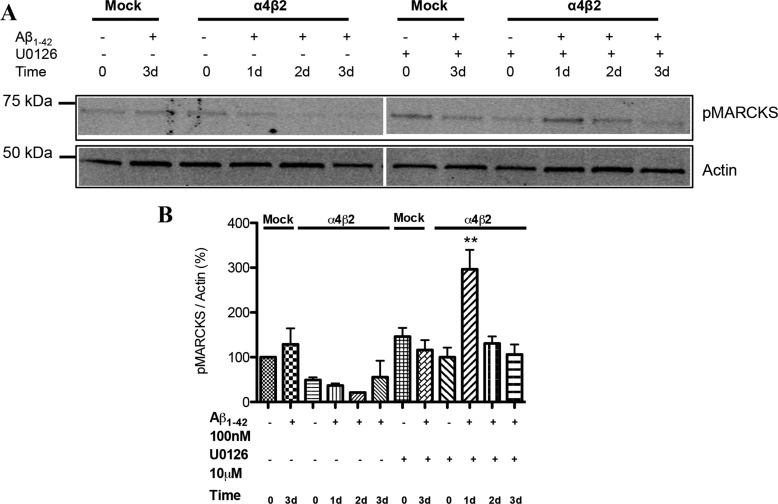
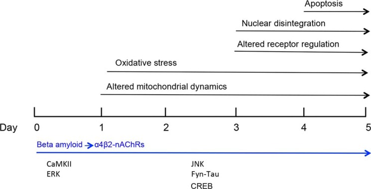
Among putative downstream synaptic targets of β-amyloid (Aβ) are signaling molecules involved in synaptic function, memory formation and cognition, such as the MAP kinases, MKPs, CaMKII, CREB, Fyn, and Tau. Here, we assessed the activation and interaction of signaling pathways upon prolonged exposure to Aβ in model nerve cells expressing nicotinic acetylcholine receptors (nAChRs). Our goal was to characterize the steps underlying sensitization of the nerve cells to neurotoxicity when Aβ-target receptors are present. Of particular focus was the connection of the activated signaling molecules to oxidative stress. Differentiated neuroblastoma cells expressing mouse α4β2-nAChRs were exposed to Aβ1-42 for intervals from 30 min to 3 days. The cells and cell-derived protein extracts were then probed for activation of signaling pathway molecules (ERK, JNK, CaMKII, CREB, MARCKS, Fyn, tau). Our results show substantial, progressive activation of ERK in response to nanomolar Aβ exposure, starting at the earliest time point. Increased ERK activation was followed by JNK activation as well as an increased expression of PHF-tau, paralleled by increased levels of reactive oxygen species (ROS). The impact of prolonged Aβ on the levels of pERK, pJNK, and ROS was attenuated by MEK-selective and JNK-selective inhibitors. In addition, the MEK inhibitor as well as a JNK inhibitor attenuated Aβ-induced nuclear fragmentation, which followed the changes in ROS levels. These results demonstrate that the presence of nAChRs sensitizes neurons to the neurotoxic action of Aβ through the timed activation of discrete intracellular signaling molecules, suggesting pathways involved in the early stages of Alzheimer disease.

Keywords: amyloid-beta (AB); c-Jun N-terminal kinase (JNK); extracellular-signal-regulated kinase (ERK); mitogen-activated protein kinase (MAPK); nicotinic acetylcholine receptors (nAChR).


Figures

FIGURE 1.
FIGURE 1.
Activation of the ERK MAPK pathway in NG108–15 cells expressing α4β2 nAChRs in response to prolonged exposure to Aβ: time-dependence and sensitivity to a MEK inhibitor. A and B, progressive increases in pERK levels in response to prolonged exposure to 100 nm1–42 in the absence or presence of U1026, a selective inhibitor of MEK, the upstream regulator of ERK, in cells expressing α4β2-nAChRs or not (Mock: mock-transfected). pERK levels were compared with total ERK as well as actin (loading control). Data are expressed as % (± S.E.) of untreated, mock-transfected controls (n = 3 experiments). C, representative images depicting immunostaining of pERK and total ERK in mock- and α4β2-nAChR-transfected cells treated with Aβ1–42 for 3 days. pERK levels in cells expressing α4β2-nAChRs and treated with Aβ were significantly different in comparison to untreated controls.
FIGURE 2.
FIGURE 2.
Activation of the JNK MAPK pathway in NG108–15 cells expressing α4β2 nAChRs in response to prolonged exposure to Aβ: time-dependence and sensitivity to JNK inhibitor. A, progressive increases in the levels of pJNK1 as well as pJNK2,3 in response to prolonged exposure to 100 nm1–42 in the absence or presence of DJNK1, a selective inhibitor of JNK, in cells expressing α4β2-nAChRs or not (Mock: mock-transfected). B, quantification of averaged immunoblot intensities for pJNKs normalized to total JNK, as compared with actin (loading control). Data are expressed as % (± S.E.) of untreated, mock-transfected controls (n = 3 experiments). C and D, expression levels of MKP-7 in differentiated NG108–15 cells treated or not with Aβ1–42. pJNK1 and pJNK2,3 levels in cells expressing α4β2-nAChRs and treated with Aβ for 2 and 3 days were significantly different in comparison to controls.
FIGURE 3.
FIGURE 3.
Interplay between ERK and JNK pathways activated in cells expressing α4β2 nAChRs in response to prolonged exposure to Aβ. A, the levels of pERK1/2 were attenuated in response to prolonged exposure to 100 nm1–42 in the presence of DJNK1, a selective inhibitor of JNK, in cells expressing α4β2-nAChRs. Similarly, levels of pJNK1 as well as pJNK2,3 were reduced significantly on incubation with U1026, MEK inhibitor. B and C represent the quantification of averaged immunoblot intensities for pERK normalized to total ERK and pJNKs normalized to total JNK, as compared with actin (loading control), respectively. *, p < 0.05 compared with controls not treated with Aβ.
FIGURE 4.
FIGURE 4.
Effects of ERK and JNK inhibitors on Aβ-induced oxidative stress in the presence of α4β2 nAChRs. A, alterations in oxidative stress (as changes in ROS) and nuclear integrity (HOESCHT staining) in differentiated NG108–15 cells expressing high-affinity nAChRs (right: α4β2) or not (left: Mock) in response to 3-day treatment with 100 nm1–42 in the absence (control) or presence of either 10 μm U0126 (MEK inhibitor) or DJNK1 (JNK inhibitor) or both. As a positive control, ROS and nuclear integrity were assessed following treatment with 100 μm hydrogen peroxide. Quantification of HOESCHT (B) and ROS (C) staining is presented as the means (± S.E.) of fluorescent intensity and number (±S.E.) of disintegrated nuclei/total nuclei (magnified example in inset), respectively, for all cells in a given field (3 experiments). Signals in cells expressing α4β2-nAChRs and treated with Aβ in presence of inhibitor were not significantly different from control (mock-transfected) cells treated with Aβ. **, p < 0.01: ***, p < 0.001 compared with controls.
FIGURE 5.
FIGURE 5.
Alterations in the levels of PHF-Tau and pFyn in response to prolonged exposure to Aβ: receptor-dependence. Progressive increases in PHF-Tau (A) and pFyn (B) in differentiated NG108–15 cells expressing α4β2-nAChRs or not (Mock: mock-transfected) response to prolonged exposure to 100 nm1–42. Note the significant increases following 3-day treatment with Aβ in the absence of target receptor (Mock). Aβ-induced increases in PHF-Tau were compared in the absence or presence of U1026, a selective inhibitor of MEK, while Aβ-induced increases in pFyn were compared in the absence or presence of PP2, a Src kinase family inhibitor. C and D, quantification of averaged immunoblot intensities, as compared with actin (loading control). Data are expressed as % (± S.E.) of untreated, mock-transfected controls (n = 3 experiments). **, p < 0.01 for PHF-tau or pFyn levels in cells expressing α4β2-nAChRs and treated with Aβ for 2 and 3 days when compared with untreated control.
FIGURE 6.
FIGURE 6.
Alterations in the levels of pCaMKII and pCREB Fyn in response to prolonged exposure to Aβ. A, progressive increases in the levels of pCaMKII and pCREB as compared with total CaMKII in differentiated NG108–15 cells expressing α4β2-nAChRs or not (Mock: mock-transfected) response to prolonged exposure to 100 nm1–42. B and C, quantification of averaged immunoblot intensities, as compared with actin (loading control) for pCaMKII and pCREB, respectively. Data are expressed as % (±S.E.) of untreated, mock-transfected controls (n = 3 experiments). *, p < 0.05 or **, p < 0.01 for pCaMKII or pCREB levels in cells expressing α4β2-nAChRs and treated with Aβ for 2 and 3 days when compared with untreated control.
FIGURE 7.
FIGURE 7.
Alterations in the levels of pMARCKS in response to prolonged exposure to Aβ: down-regulation and the impact of ERK inhibition. A, progressive decreases in the levels of pMARCKS in differentiated NG108–15 cells expressing α4β2-nAChRs or not (Mock: mock-transfected) response to prolonged exposure to 100 nm1–42 in the absence (control) or presence of 10 μm U0126 (MEK inhibitor). Progressive decreases in the levels of Western blot micrograph showing the altered levels of pCREB and pCaMKII in mock and α4β2-nAChR-transfected cells treated with Aβ for different time-points. GAPDH was used as a loading control. B and C show the quantification of the blots for pCREB and pCaMKII respectively. Data are expressed as % (±S.E.) of untreated, mock-transfected controls (n = 3 experiments). **, p < 0.01 compared with controls.
FIGURE 8.
FIGURE 8.
Schematic timeline for sensitization of Aβ-induced neurotoxicity in the presence of high-affinity nAChRs: Steps leading to apoptosis (22) and the key signaling molecules involved.

References

    1. Holtzman D. M., Morris J. C., Goate A. M. (2011) Alzheimer's disease: the challenge of the second century. Sci. Transl. Med. 3, 77sr1. - PMC - PubMed
    1. Balleza-Tapia H., Peña F. (2009) Pharmacology of the intracellular pathways activated by Amyloid beta protein. Mini-Rev. Med. Chem. 9, 724–740 - PubMed
    1. Pei J. J., Braak E., Braak H., Grundke-Iqbal I., Iqbal K., Winblad B., Cowburn R. F. (2001) Localization of active forms of C-jun kinase (JNK) and p38 kinase in Alzheimer's disease brains at different stages of neurofibrillary degeneration. J. Alzheimers Dis. 3, 41–48 - PubMed
    1. Ferrer I., Blanco R., Carmona M., Ribera R., Goutan E., Puig B., Rey M. J., Cardozo A., Viñals R., Ribalta T. (2001) Phosphorylated MAP kinase (ERK1, ERK2) expression is associated with early tau deposition in neurons and glial cells, but not with increased nuclear DNA vulnerability and cell death, in Alzheimer's disease, Pick's disease, progressive supranuclear palsy and corticobasal degeneration. Brain Pathol. 11, 144–158 - PMC - PubMed
    1. Dineley K. T., Westerman M., Bui D., Bell K., Ashe K. H., Sweatt J. D. (2001) Beta-amyloid activates the mitogen-activated protein kinase cascade via hippocampal alpha7 nicotinic acetylcholine receptors: In vitro and in vivo mechanisms related to Alzheimer's disease. J. Neurosci. 21, 4125–4133 - PMC - PubMed

Publication types