

Use of magnetic resonance elastography for assessing liver functional reserve: A clinical study
- PMID: 26139999
- PMCID: PMC4481448
- DOI: 10.3748/wjg.v21.i24.7522
Use of magnetic resonance elastography for assessing liver functional reserve: A clinical study
Abstract
Aim: To investigate the value of magnetic resonance elastography (MRE) with regard to assessing liver functional reserve.
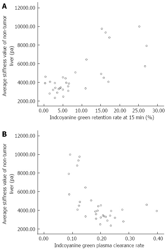
Methods: Data from inpatients diagnosed with a liver tumor at an interventional radiology department from July 2013 to June 2014 were analyzed. A 3.0 Tesla magnetic resonance unit was used to scan 32 patients with confirmed diagnoses of hepatocellular carcinoma (HCC); an MRE sequence was added to the protocol, and the data were reconstructed and analyzed by two attending radiologists. Regions of interest were identified in different slices of the non-tumor liver parenchyma to measure average stiffness. In addition, the indocyanine green (ICG) test was performed no more than 1 wk before or after the magnetic resonance examination for all 32 patients; the ICG retention rate at 15 min (ICGR-15) and the ICG plasma clearance rate (ICG-K) were recorded. Correlational analyses were performed between the liver stiffness values and the ICGR-15 as well as between the liver stiffness values and the ICG-K.
Results: Magnetic resonance imaging, including an MRE sequence and the ICG test, was performed successfully in all 32 enrolled patients. None of the patients developed complications. The mean ± SD of the elasticity values measured by the two attending radiologists were 4.7 ± 2.2 kPa and 4.7 ± 2.1 kPa, respectively. The average liver stiffness value of the non-tumor parenchyma measured using MRE in HCC patients was 4.7 ± 2.2 kPa. The average ICGR-15 was 0.089 ± 0.077, and the average ICG-K was 0.19 ± 0.07. We found that the liver stiffness value of the non-tumor parenchyma was significantly and positively related to the ICGR-15 (r = 0.746, P < 0.01) as well as significantly and negatively related to the ICG-K (r = -0.599, P < 0.01). The ICGR-15 was significantly and negatively related to the ICG-K (r = -0.852, P < 0.01).
Conclusion: MRE is accurate and non-invasive; furthermore, it can be used to effectively assess the liver functional reserve of HCC patients.
Keywords: Hepatocellular carcinoma; Indocyanine green clearance test; Liver fibrosis; Liver functional reserve; Magnetic resonance elastography.
Figures
References
-
- Llovet JM, Beaugrand M. Hepatocellular carcinoma: present status and future prospects. J Hepatol. 2003;38 Suppl 1:S136–S149. - PubMed
-
- Poon RT, Fan ST. Assessment of hepatic reserve for indication of hepatic resection: how I do it. J Hepatobiliary Pancreat Surg. 2005;12:31–37. - PubMed
-
- Mizuguchi T, Kawamoto M, Meguro M, Hui TT, Hirata K. Preoperative liver function assessments to estimate the prognosis and safety of liver resections. Surg Today. 2014;44:1–10. - PubMed
-
- Chen X, Zhang HB, Li ZQ, Yu XF, Yang MF, Wang HH, Teng LS. Indocyanine green clearance in evaluating the recovery of liver reserve function after superselective transarterial chemoembolization. Hepatobiliary Pancreat Dis Int. 2013;12:656–660. - PubMed
Publication types
MeSH terms
Substances
Associated data
LinkOut - more resources
Full Text Sources
Other Literature Sources
Medical
