

Case Reports
doi: 10.4250/jcu.2015.23.2.119.
Epub 2015 Jun 26.
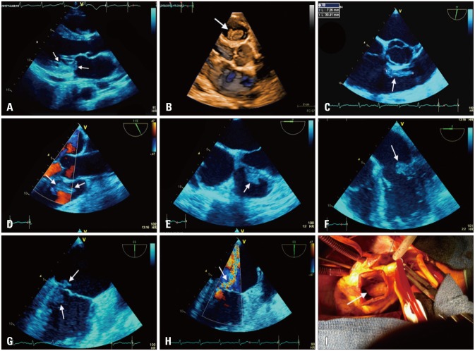
Simultaneous Bilateral Infective Endocarditis with Right Ventricular Mural Involvement
Affiliations
- PMID: 26140157
- PMCID: PMC4486178
- DOI: 10.4250/jcu.2015.23.2.119
Item in Clipboard
Case Reports
Simultaneous Bilateral Infective Endocarditis with Right Ventricular Mural Involvement
J Cardiovasc Ultrasound.
2015 Jun.
No abstract available
Keywords: Cardiac surgery; Echocardiography; Endocarditis; Infective endocarditis.
Figures
References
-
- Yao F, Han L, Xu ZY, Zou LJ, Huang SD, Wang ZN, Lu FL, Yao YL. Surgical treatment of multivalvular endocarditis: twenty-one-year single center experience. J Thorac Cardiovasc Surg. 2009;137:1475–1480. - PubMed
Publication types
LinkOut - more resources
Full Text Sources
Other Literature Sources
