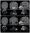
Intraoperative fiducial-less patient registration using volumetric 3D ultrasound: a prospective series of 32 neurosurgical cases
- PMID: 26140481
- PMCID: PMC4778720
- DOI: 10.3171/2014.12.JNS141321
Intraoperative fiducial-less patient registration using volumetric 3D ultrasound: a prospective series of 32 neurosurgical cases
Abstract
Object: Fiducial-based registration (FBR) is used widely for patient registration in image-guided neurosurgery. The authors of this study have developed an automatic fiducial-less registration (FLR) technique to find the patient-to-image transformation by directly registering 3D ultrasound (3DUS) with MR images without incorporating prior information. The purpose of the study was to evaluate the performance of the FLR technique when used prospectively in the operating room and to compare it with conventional FBR.
Methods: In 32 surgical patients who underwent conventional FBR, preoperative T1-weighted MR images (pMR) with attached fiducial markers were acquired prior to surgery. After craniotomy but before dural opening, a set of 3DUS images of the brain volume was acquired. A 2-step registration process was executed immediately after image acquisition: 1) the cortical surfaces from pMR and 3DUS were segmented, and a multistart sum-of-squared-intensity-difference registration was executed to find an initial alignment between down-sampled binary pMR and 3DUS volumes; and 2) the alignment was further refined by a mutual information-based registration between full-resolution grayscale pMR and 3DUS images, and a patient-to-image transformation was subsequently extracted.
Results: To assess the accuracy of the FLR technique, the following were quantified: 1) the fiducial distance error (FDE); and 2) the target registration error (TRE) at anterior commissure and posterior commissure locations; these were compared with conventional FBR. The results showed that although the average FDE (6.42 ± 2.05 mm) was higher than the fiducial registration error (FRE) from FBR (3.42 ± 1.37 mm), the overall TRE of FLR (2.51 ± 0.93 mm) was lower than that of FBR (5.48 ± 1.81 mm). The results agreed with the intent of the 2 registration techniques: FBR is designed to minimize the FRE, whereas FLR is designed to optimize feature alignment and hence minimize TRE. The overall computational cost of FLR was approximately 4-5 minutes and minimal user interaction was required.
Conclusions: Because the FLR method directly registers 3DUS with MR by matching internal image features, it proved to be more accurate than FBR in terms of TRE in the 32 patients evaluated in this study. The overall efficiency of FLR in terms of the time and personnel involved is also improved relative to FBR in the operating room, and the method does not require additional image scans immediately prior to surgery. The performance of FLR and these results suggest potential for broad clinical application.
Keywords: 3DUS = 3D ultrasound; 3DUS-MR registration; AC = anterior commissure; FBR = fiducial-based registration; FDE = fiducial distance error; FLR = fiducial-less registration; FRE = fiducial registration error; OR = operating room; PC = posterior commissure; SBR = surface-based registration; TRE = target registration error; diagnostic and operative techniques; image-guided neurosurgery; pMR = preoperative MR images; patient registration; volumetric 3D ultrasound.
Figures


Similar articles
-
Intraoperative patient registration using volumetric true 3D ultrasound without fiducials.Med Phys. 2012 Dec;39(12):7540-52. doi: 10.1118/1.4767758. Med Phys. 2012. PMID: 23231302 Free PMC article.
-
Intraoperative image updating for brain shift following dural opening.J Neurosurg. 2017 Jun;126(6):1924-1933. doi: 10.3171/2016.6.JNS152953. Epub 2016 Sep 9. J Neurosurg. 2017. PMID: 27611206 Free PMC article.
-
Registration of CT to 3D ultrasound using near-field fiducial localization: A feasibility study.Comput Aided Surg. 2011;16(2):54-70. doi: 10.3109/10929088.2011.556181. Comput Aided Surg. 2011. PMID: 21322745
-
IBIS: an OR ready open-source platform for image-guided neurosurgery.Int J Comput Assist Radiol Surg. 2017 Mar;12(3):363-378. doi: 10.1007/s11548-016-1478-0. Epub 2016 Aug 31. Int J Comput Assist Radiol Surg. 2017. PMID: 27581336 Review.
-
Multimodal 3D ultrasound and CT in image-guided spinal surgery: public database and new registration algorithms.Int J Comput Assist Radiol Surg. 2021 Apr;16(4):555-565. doi: 10.1007/s11548-021-02323-2. Epub 2021 Mar 8. Int J Comput Assist Radiol Surg. 2021. PMID: 33683544 Review.
Cited by
-
Surgical Navigation System for Hypertensive Intracerebral Hemorrhage Based on Mixed Reality.J Digit Imaging. 2022 Dec;35(6):1530-1543. doi: 10.1007/s10278-022-00676-x. Epub 2022 Jul 11. J Digit Imaging. 2022. PMID: 35819536 Free PMC article.
-
Regional-surface-based registration for image-guided neurosurgery: effects of scan modes on registration accuracy.Int J Comput Assist Radiol Surg. 2019 Aug;14(8):1303-1315. doi: 10.1007/s11548-019-01990-6. Epub 2019 May 4. Int J Comput Assist Radiol Surg. 2019. PMID: 31055765
-
Applications of Ultrasound in the Resection of Brain Tumors.J Neuroimaging. 2017 Jan;27(1):5-15. doi: 10.1111/jon.12382. Epub 2016 Aug 19. J Neuroimaging. 2017. PMID: 27541694 Free PMC article. Review.
-
3D intra-operative ultrasound and MR image guidance: pursuing an ultrasound-based management of brainshift to enhance neuronavigation.Int J Comput Assist Radiol Surg. 2017 Oct;12(10):1711-1725. doi: 10.1007/s11548-017-1578-5. Epub 2017 Apr 8. Int J Comput Assist Radiol Surg. 2017. PMID: 28391583
References
-
- Buckner JC. Factors influencing survival in high-grade gliomas. Semin Oncol. 2003;30(6 Suppl 19):10–14. - PubMed
-
- Carter TJ, Sermesant M, Cash DM, Barratt DC, Tanner C, Hawkes DJ. Application of soft tissue modelling to image-guided surgery. Med Eng Phys. 2005;27:893–909. - PubMed
-
- Claus EB, Horlacher A, Hsu L, Schwartz RB, Dello-Iacono D, Talos F, et al. Survival rates in patients with low-grade glioma after intraoperative magnetic resonance image guidance. Cancer. 2005;103:1227–1233. - PubMed
-
- Fan X, Ji S, Fontaine K, Hartov A, Roberts D, Paulsen K. Simulation of brain tumor resection in image-guided neurosurgery. Proc SPIE. 2011;7964:79640U.
Publication types
MeSH terms
Grants and funding
LinkOut - more resources
Full Text Sources
Other Literature Sources

