. 2015 Jul 2;162(1):134-45.
doi: 10.1016/j.cell.2015.06.027.

Neural Representations of Unconditioned Stimuli in Basolateral Amygdala Mediate Innate and Learned Responses

Affiliations

Neural Representations of Unconditioned Stimuli in Basolateral Amygdala Mediate Innate and Learned Responses

Felicity Gore et al. Cell. .

Abstract

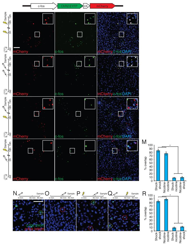
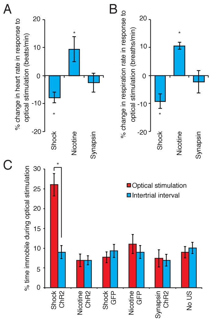
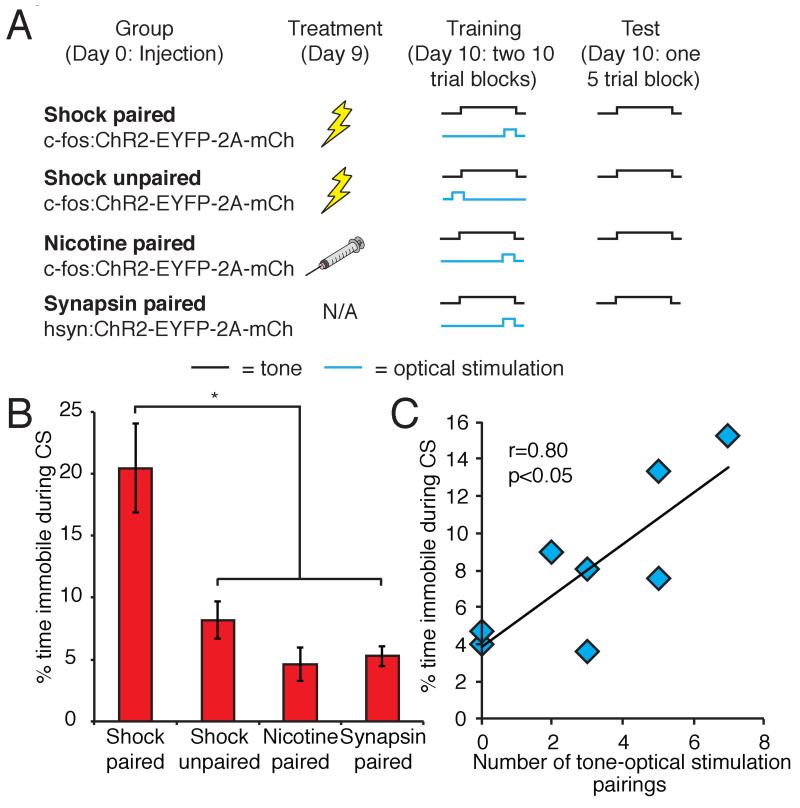
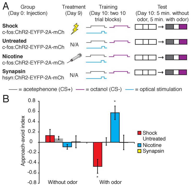
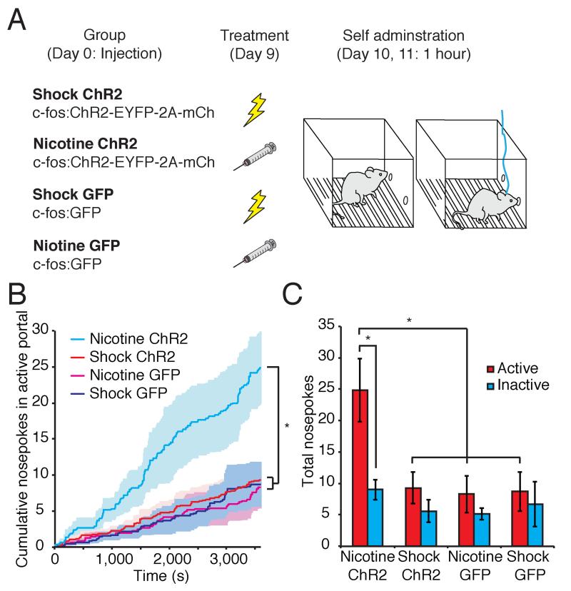
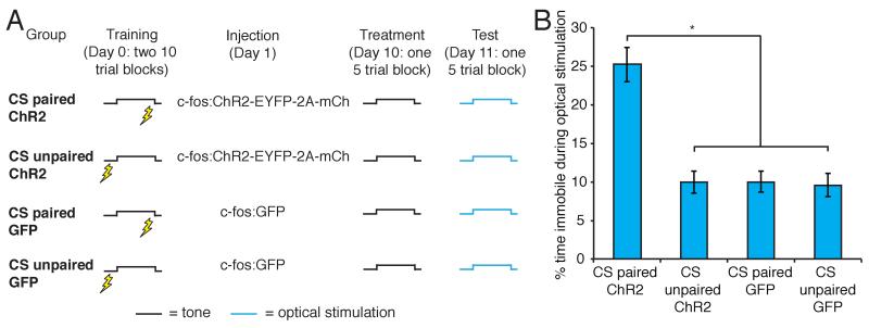
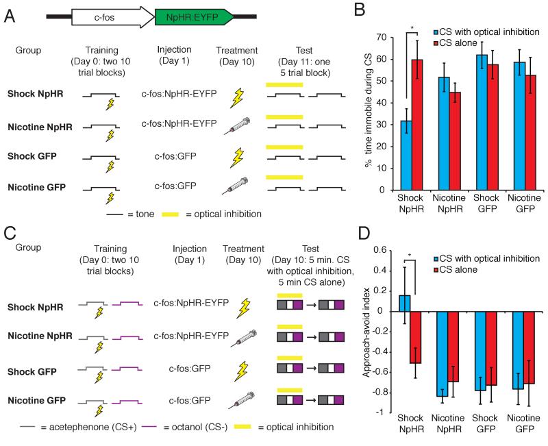
Stimuli that possess inherently rewarding or aversive qualities elicit emotional responses and also induce learning by imparting valence upon neutral sensory cues. Evidence has accumulated implicating the amygdala as a critical structure in mediating these processes. We have developed a genetic strategy to identify the representations of rewarding and aversive unconditioned stimuli (USs) in the basolateral amygdala (BLA) and have examined their role in innate and learned responses. Activation of an ensemble of US-responsive cells in the BLA elicits innate physiological and behavioral responses of different valence. Activation of this US ensemble can also reinforce appetitive and aversive learning when paired with differing neutral stimuli. Moreover, we establish that the activation of US-responsive cells in the BLA is necessary for the expression of a conditioned response. Neural representations of conditioned and unconditioned stimuli therefore ultimately connect to US-responsive cells in the BLA to elicit both innate and learned responses.


Figures

Figure 1
Figure 1. Anatomically distinct, yet intermingled populations of cells in the BLA respond to appetitive and aversive unconditioned stimuli
A-C: Animals injected with lentivirus expressing c-fos:ChR2-EYFP-2A-mCherry were exposed to 2 footshock treatments separated by 18 hours and stained for mCherry (A), c-fos (B), and merged (C). D-F: Animals injected with lentivirus expressing c-fos:ChR2-EYFP-2A-mCherry were treated with 2 i.p. nicotine injections separated by 18 hours and stained for mCherry (D), c-fos (E), and merged (F). G-I: Animals injected with lentivirus expressing c-fos:ChR2-EYFP-2A-mCherry were treated with footshock followed by nicotine 18 hours later and stained for mCherry (G), c-fos (H), and merged (I). J-L: Animals injected with lentivirus expressing c-fos:ChR2-EYFP-2A-mCherry were treated with nicotine followed by footshock 18 hours later and stained for mCherry (J), and c-fos (K), and merged (L). M. Percent overlap ((mCherry+ + c-fos+)/mCherry+) of c-fos positive and mCherry positive neurons in the BLA (shock-shock 84.07±4.46, n=6; nicotine-nicotine 76.02±4.90, n=5; shock-nicotine 8.22±1.40, n=6; nicotine-shock 9.28±2.94, n=5. One-way ANOVA, F3,18=130.43, P<0.0001). N-Q: N. Animals exposed to nicotine treatment 45 minutes prior to sacrifice and stained for intronic c-fos RNA (nuclear, red) and exonic c-fos RNA (cytoplasmic, green). O. Animals exposed to nicotine treatment 5 minutes prior to sacrifice and stained for intronic c-fos RNA and exonic c-fos RNA. P. Animals exposed to footshock treatment 45 minutes prior to sacrifice and nicotine treatment 5 minutes prior to sacrifice and stained for intronic c-fos RNA and exonic c-fos RNA. Q. Animals exposed to nicotine treatment 45 minutes prior to sacrifice and footshock treatment 5 minutes prior to sacrifice and stained for intronic c-fos RNA and exonic c-fos RNA. R. Percent overlap (yellow/green) of c-fos intronic RNA positive neurons (nuclear red) with c-fos exonic RNA positive neurons (cytoplasmic green) (shock-shock 82.19±3.86%, n=3; nicotine-nicotine 87.93±3.29%, n=4; shock-nicotine 8.63±2.67%, n=4; nicotine-shock 11.62±1.75%, n=6. One-way ANOVA, F3,13=254.29, P<0.0001). Scale bars, 100μm. All error bars for all figures represent ± s.e.m. See also Figure S2.
Figure 2
Figure 2. The exogenous activation of cells responsive to footshock and nicotine in the BLA is sufficient to elicit valence-specific physiologic and behavioral responses
A. Percent change in heart rate from baseline in response to optical stimulation of footshock or nicotine responsive cells, or a random ensemble (shock −7.88±1.93% n=5; nicotine 9.49±4.52% n=5; synapsin −2.55±3.40%, n=6. Two-way ANOVA, group × optical stimulation interaction, F2,26=6.24, P<0.01). B. Percent change in respiration rate from baseline in response to optical stimulation of footshock or nicotine responsive cells, or a random ensemble (shock −9.20±2.55%, n=5; nicotine 10.63±1.21%, n=5; synapsin - 2.19±4.05%, n=6. Two-way ANOVA, group × optical stimulation interaction, F2,26=10.14, P<0.001). C. Percent of time spent freezing in response to optical stimulation compared to the intertrial interval (ITI) (shock ChR2 optical stimulation 26.05±2.83%, ITI 8.99±1.66% n=7; nicotine ChR2 optical stimulation 6.93±1.58%, ITI 6.94±1.44 n=6; shock GFP optical stimulation 7.70±1.37%, ITI 9.32±1.65, n=6; nicotine GFP optical stimulation 11.07±2.42%, ITI 9.02±1.62%, n=6; synapsin ChR2 optical stimulation 7.43±2.19%, ITI 6.95±1.46%, n=5; no US optical stimulation 8.96±1.42%, ITI 10.05±1.40, n=5. Two-way ANOVA, group × optical stimulation interaction, F5,58=8.28, P<0.0001). See also Figures S3 and S4.
Figure 3
Figure 3. The exogenous activation of footshock-responsive cells can act as an US in an auditory fear conditioning paradigm
A. Modified fear conditioning paradigm. B. Percent time spent freezing in response to the auditory CS following fear conditioning using optical stimulation of the BLA as the US (shock paired 20.46±3.59%, n=10; shock unpaired 8.19±1.51%, n=8; nicotine paired 4.62±1.38%, n=8, synapsin paired 5.26±0.80%, n=6. One-way ANOVA, F3,28=9.74, P<0.0005). C. Correlation between number of tone-optical stimulation pairings and percent immobility to CS in shock unpaired animals.
Figure 4
Figure 4. The exogenous activation of footshock- and nicotine-responsive cells can reinforce aversive and appetitive olfactory conditioning, respectively
A. Behavioral training protocol for associative olfactory learning. B. Approach-avoid index (difference in time spent in CS+ and CS-compartments of a 3 compartment chamber, divided by the time spent in both compartments) of shock-induced, untreated and nicotine-induced animals trained to associate odor with optical stimulation of the BLA, as well as animals expressing ChR2 in a random population of BLA neurons (shock without odor 0.13±0.12, with odor −0.47±0.14, n=6; untreated without odor 0.08±0.03, with odor 0.02±0.07, n=6; nicotine without odor −0.09±0.05, with odor 0.57±0.13, n=6; synapsin without odor 0.01±0.12, with odor 0.00±0.14, n=5. Two-way ANOVA, group × conditioning interaction, F3,38=12.65, P<0.0005).
Figure 5
Figure 5. The exogenous activation of nicotine-responsive cells can reinforce instrumental conditioning
A. Behavioral protocol for instrumental conditioning B. Average cumulative nosepokes for the active portal on the second day of testing in animals expressing ChR2 or GFP in footshock or nicotine responsive cells. Shading represents ± s.e.m. (One-way ANOVA, F3,26=5.08, P<0.01). C. Total nosepokes in the active and inactive portal on the second day of testing (nicotine-ChR2 active 24.83±5.02, inactive 9.00±1.57, n=6; shock-ChR2 active 9.30±2.54, inactive 5.60±1.81, n=10; nicotine-GFP active 8.29±2.92, inactive 5.14±0.91, n=7; shock-GFP active 8.71±3.11, inactive 6.71±3.58, n=7. Two-way ANOVA, group F3,52=5.08, P<0.01, portal F1,52=8.03, P<0.01).
Figure 6
Figure 6. The exogenous activation of a learned aversive CS representation can drive freezing behavior, whereas the exogenous activation of an unlearned CS representation cannot
A. Behavioral protocol for the reactivation of learned and unlearned CS representations in the BLA. B. Percent of time spent freezing in response to optical stimulation of the CS representation in the BLA (CS paired ChR2 25.22±2.25%, n=9; CS unpaired ChR2 9.93±1.40%, n=10; CS paired GFP 9.98±1.36%, n=6; CS unpaired GFP 9.59±1.50%, n=6. One-way ANOVA, F3,26=20.10, P<0.00001).
Figure 7
Figure 7. Learning connects auditory and olfactory CS representations to US-responsive neurons in the BLA
A. Behavioral protocol for the silencing of US-responsive cells during auditory CS presentation. B. Percent immobility in response to the CS in the presence and absence of optical inhibition of footshock and nicotine-responsive cells (shock NpHR with yellow light 31.75±5.62%, without yellow light 59.84±8.72%, n=6; nicotine NpHR with yellow light 51.70±6.54%, without yellow light 44.73%±4.45%, n=9; shock GFP with yellow light 61.92±6.06%, without yellow light 57.59±6.55%, n=6; nicotine GFP with yellow light 58.59±5.84%, without yellow light 52.64±8.18%, n=6. Two-way ANOVA, group × optical inhibition interaction, F3,46=3.16, P<0.05). C. Behavioral protocol for the silencing of US-responsive cells during olfactory CS presentation. D. Approach-avoid index in the presence and absence of optical inhibition of footshock and nicotine-responsive cells (shock NpHR with yellow light 0.16±0.28, without yellow light −0.51±0.15, n=6; nicotine NpHR with yellow light - 0.83±0.07, without yellow light −0.69±0.15, n=6; shock GFP with yellow light - 0.78±0.13, without yellow light, −0.72±0.17, n=6; nicotine GFP with yellow light - 0.76±0.15, without yellow light −0.71±0.22, n=6. Two-way ANOVA, group F3,40=5.36, P<0.005). See also Figures S5 and S6.

Comment in

References

    1. Amano T, Duvarci S, Popa D, Paré D. The fear circuit revisited: contributions of the basal amygdala nuclei to conditioned fear. J Neurosci. 2011;31:15481–15489. - PMC - PubMed
    1. Amaral D, Price J, Pitkanen A, Carmichael S. In: The Amygdala: Neurobiological Aspects of Emotion, Memory, and Mental Dysfunction. Aggleton J, editor. Wiley-Liss; New York: 1992. pp. 1–66.
    1. Ambroggi F, Ishikawa A, Fields HL, Nicola SM. Basolateral amygdala neurons facilitate reward-seeking behavior by exciting nucleus accumbens neurons. Neuron. 2008;59:648–661. - PMC - PubMed
    1. Anglada-Figueroa D, Quirk GJ. Lesions of the basal amygdala block expression of conditioned fear but not extinction. J Neurosci. 2005;25:9680–9685. - PMC - PubMed
    1. Balleine BW, Killcross S. Parallel incentive processing: an integrated view of amygdala function. Trends Neurosci. 2006;29:272–279. - PubMed

Publication types