. 2016 Feb;173(4):766-77.
doi: 10.1111/bph.13237. Epub 2015 Jul 31.

Neutrophil elastase induces inflammation and pain in mouse knee joints via activation of proteinase-activated receptor-2

Affiliations

Neutrophil elastase induces inflammation and pain in mouse knee joints via activation of proteinase-activated receptor-2

Milind M Muley et al. Br J Pharmacol. 2016 Feb.

Abstract

Background and purpose: Neutrophil elastase plays a crucial role in arthritis. Here, its potential in triggering joint inflammation and pain was assessed, and whether these effects were mediated by proteinase-activated receptor-2 (PAR2).

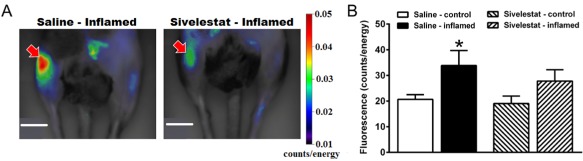
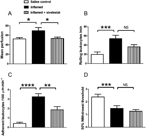
Experimental approach: Neutrophil elastase (5 μg) was injected into the knee joints of mice and changes in blood perfusion, leukocyte kinetics and paw withdrawal threshold were assessed. Similar experiments were performed in animals pretreated with the neutrophil elastase inhibitor sivelestat, the PAR2 antagonist GB83, the p44/42 MAPK inhibitor U0126 and in PAR2 receptor knockout (KO) mice. Neutrophil elastase activity was also evaluated in arthritic joints by fluorescent imaging and sivelestat was assessed for anti-inflammatory and analgesic properties.

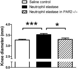
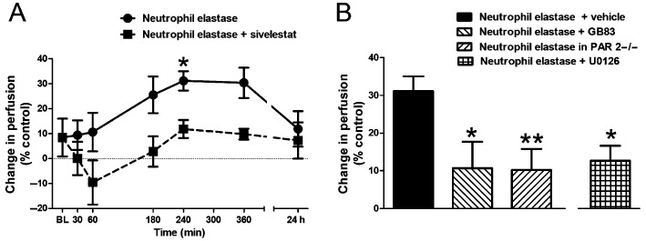
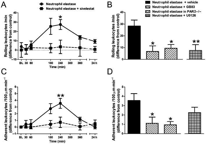
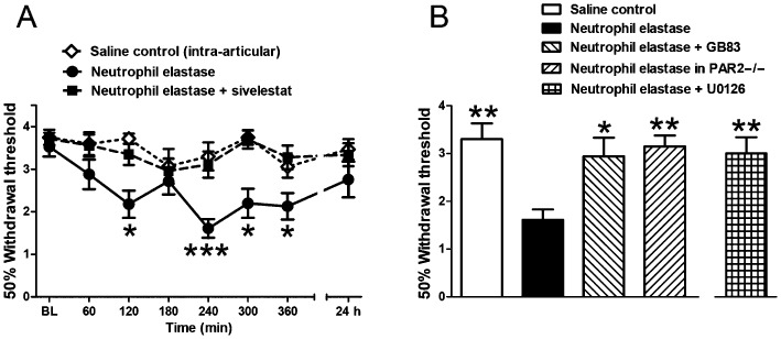
Key results: Intra-articular injection of neutrophil elastase caused an increase in blood perfusion, leukocyte kinetics and a decrease in paw withdrawal threshold. Sivelestat treatment suppressed this effect. The PAR2 antagonist GB83 reversed neutrophil elastase-induced synovitis and pain and these responses were also attenuated in PAR2 KO mice. The MAPK inhibitor U0126 also blocked neutrophil elastase-induced inflammation and pain. Active neutrophil elastase was increased in acutely inflamed knees as shown by an activatable fluorescent probe. Sivelestat appeared to reduce neutrophil elastase activity, but had only a moderate anti-inflammatory effect in this model.

Conclusions and implications: Neutrophil elastase induced acute inflammation and pain in knee joints of mice. These changes are PAR2-dependent and appear to involve activation of a p44/42 MAPK pathway. Blocking neutrophil elastase, PAR2 and p44/42 MAPK activity can reduce inflammation and pain, suggesting their utility as therapeutic targets.


Figures

Figure 1
Figure 1
Knee diameter in mice. Knee joint diameters, measured 4 h following intra‐articular injection of neutrophil elastase, showing a significant increase in wild‐type mice, but not in PAR2 KO mice (PAR 2−/−), when compared with intra‐articular saline. Values represent mean ± SEM (n = 9–15/group). *P < 0.05, ***P < 0.001 compared with neutrophil elastase.
Figure 2
Figure 2
Changes in vascular perfusion. (A) Time course of the increase in knee joint blood perfusion following intra‐articular injection of neutrophil elastase and its reversal by systemic pretreatment with the neutrophil elastase inhibitor sivelestat (n = 6–7 per time point). *P < 0.05 compared with baseline (BL). (B) Mean perfusion in the knee joint microvasculature at 4 h following intra‐articular injection of neutrophil elastase showing the increased perfusion is blocked by the PAR2 antagonist GB83 and is absent in PAR2 KO mice (PAR 2/). The neutrophil elastase effect is also blocked by the MAPK inhibitor U0126. Values shown are the per cent difference in perfusion units compared with the saline‐injected contralateral knee and are means ± SEM (n = 9–15/group). *P < 0.05, **P < 0.01 compared with neutrophil elastase alone.
Figure 3
Figure 3
Changes in leukocyte trafficking. Time course of the increase in the number of (A) rolling and (C) adherent leukocytes following intra‐articular injection of neutrophil elastase and its reversal by systemic pretreatment with sivelestat (n = 6–7 per time point). *P < 0.05, **P < 0.01 compared with baseline (BL). Increases in the number of (B) rolling and (D) adherent leukocytes in the knee joint microvasculature at 4 h following intra‐articular injection of neutrophil elastase are blocked by the PAR2 antagonist GB83 and are absent in PAR2 KO mice (PAR 2/). The effect of neutrophil elastase on rolling leukocytes is also blocked by the MAPK inhibitor U0126. Values shown are the difference in number of cells compared with the saline‐injected contralateral knee and are means ± SEM (n = 9–15/group). *P < 0.05, **P < 0.01 compared with neutrophil elastase alone.
Figure 4
Figure 4
Changes in behavioural pain. (A) Time course of the decrease in withdrawal threshold following intra‐articular neutrophil elastase and its reversal by systemic pretreatment with the neutrophil elastase inhibitor sivelestat. The intra‐articular saline control did not show a change in withdrawal threshold. *P < 0.05, ***P < 0.001 compared with baseline (BL). (B) Withdrawal thresholds at 4 h following intra‐articular neutrophil elastase showing the induced change is blocked by pretreatment with the PAR2 antagonist GB83 and is absent in PAR2 KO mice (PAR 2/). The neutrophil elastase effect is also blocked by the MAPK inhibitor U0126. Values shown are von Frey 50% withdrawal thresholds and are mean ± SEM (n = 9–11/group). *P < 0.05, **P < 0.01 compared with neutrophil elastase alone.
Figure 5
Figure 5
In vivo imaging of neutrophil elastase enzyme activity. (A) Representative fluorescence images taken 24 h after unilateral induction of knee joint inflammation with kaolin‐carrageenan (right knee, indicated by arrow) compared with the untreated contralateral joint (left knee). Arthritic animals were treated with either systemic saline (left image) or systemic sivelestat (right image). Sivelestat significantly reduced neutrophil elastase activity in inflamed joints (arrow in right image). Scale bar = 1 cm. (B) Fluorescence intensity in the inflamed and control knee joints. Values shown are means ± SEM (n = 5–6 per group). *P < 0.05 compared with saline control.
Figure 6
Figure 6
Kaolin/carrageenan‐induced inflammation and pain. Changes in (A) mean perfusion, (B) rolling and (C) adherent leukocytes, and (D) secondary allodynia in the knee joint 24 h following intra‐articular administration of saline (control), or kaolin‐carrageenan (inflamed) with or without systemic administration of sivelestat. Sivelestat treatment significantly inhibited joint inflammation but not joint pain. Values shown are means ± SEM (n = 10–12 per group). NS = not significant, *P < 0.05, **P < 0.01, ***P < 0.001, ****P < 0.0001 compared with inflamed control.

References

    1. Adeyemi EO, Campos LB, Loizou S, Walport MJ, Hodgson HJF (1990). Plasma lactoferrin and neutrophil elastase in rheumatoid arthritis and systemic lupus erythematosus. Br J Rheumatol 29: 15–20. - PubMed
    1. Alexander SPH, Benson HE, Faccenda E, Pawson AJ, Sharman JL, Spedding M et al (2013a). The concise guide to PHARMACOLOGY 2013/14: G protein‐coupled receptors. Br J Pharmacol 170: 1459–1581. - PMC - PubMed
    1. Alexander SPH, Benson HE, Faccenda E, Pawson AJ, Sharman JL, Catterall WA et al (2013b). The Concise Guide to PHARMACOLOGY 2013/14: Ion Channels. Br J Pharmacol 170: 1607–1651. - PMC - PubMed
    1. Alexander SPH, Benson HE, Faccenda E, Pawson AJ, Sharman JL, Spedding M et al (2013c). The Concise Guide to PHARMACOLOGY 2013/14: Enzymes. Br J Pharmacol 170: 1797–1867. - PMC - PubMed
    1. Andruski B, McCafferty DM, Ignacy T, Millen B, McDougall JJ (2008). Leukocyte trafficking and pain behavioral responses to a hydrogen sulfide donor in acute monoarthritis. Am J Physiol Regul Integr Comp Physiol 295: R814–R820. - PubMed

Publication types