. 2015 Jul 4:3:41.
doi: 10.1186/s40478-015-0217-z.

Impact of amyloid β aggregate maturation on antibody treatment in APP23 mice

Affiliations

Impact of amyloid β aggregate maturation on antibody treatment in APP23 mice

Karthikeyan Balakrishnan et al. Acta Neuropathol Commun. .

Abstract

Introduction: The deposition of the amyloid β protein (Aβ) in the brain is a hallmark of Alzheimer's disease (AD). Removal of Aβ by Aβ-antibody treatment has been developed as a potential treatment strategy against AD. First clinical trials showed neither a stop nor a reduction of disease progression. Recently, we have shown that the formation of soluble and insoluble Aβ aggregates in the human brain follows a hierarchical sequence of three biochemical maturation stages (B-Aβ stages). To test the impact of the B-Aβ stage on Aβ immunotherapy, we treated transgenic mice expressing human amyloid precursor protein (APP) carrying the Swedish mutation (KM670/671NL; APP23) with the Aβ-antibody β1 or phosphate-buffered saline (PBS) beginning 1) at 3 months, before the onset of dendrite degeneration and plaque deposition, and 2) at 7 months, after the start of Aβ plaque deposition and dendrite degeneration.

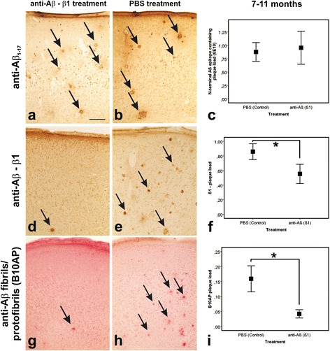
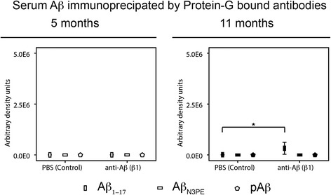
Results: At 5 months of age, first Aβ aggregates in APP23 brain consisted of non-modified Aβ (representing B-Aβ stage 1) whereas mature Aβ-aggregates containing N-terminal truncated, pyroglutamate-modified AβN3pE and phosphorylated Aβ (representing B-Aβ stage 3) were found at 11 months of age in both β1- and PBS-treated animals. Protective effects on commissural neurons with highly ramified dendritic trees were observed only in 3-month-old β1-treated animals sacrificed at 5 months. When treatment started at 7 months of age, no differences in the numbers of healthy commissural neurons were observed between β1- and PBS-treated APP23 mice sacrificed with 11 months.

Conclusions: Aβ antibody treatment was capable of protecting neurons from dendritic degeneration as long as Aβ aggregation was absent or represented B-Aβ stage 1 but had no protective or curative effect in later stages with mature Aβ aggregates (B-Aβ stage 3). These data indicate that the maturation stage of Aβ aggregates has impact on potential treatment effects in APP23 mice.


Figures

Fig. 1
Fig. 1
Degeneration of commissural neurons with a highly ramified dendritic tree: Effects of β1 antibody treatment. a, b Layer III type 1 commissural neurons in the frontocentral cortex traced with DiI. The detectable type 1 commissural neurons did not show significant differences between PBS- and β1-treated APP23 mice even at 5 months of age. c The number of the type 1 commissural neurons in 5-month-old β1 treated mice was higher than that in PBS-treated animals whereas no such differences were observed for type 2 and 3 commissural neurons. d No differences in the numbers of DiI-traced type 1–3 commissural neurons were seen among 11-month-old PBS- and β1-treated APP23 mice. Graphs represent mean values (symbols) and standard errors (whiskers), * p < 0.05 (Statistical analysis in Additional file 3: Table S3). Calibration bar in b (valid for a, b) = 37.5 μm
Fig. 2
Fig. 2
Aβ plaque pathology in the frontocentral cortex of APP23 mice: Effects of β1 antibody treatment. Aβ plaque pathology in PBS- and β1-treated APP23 mice. The staining pattern in 11-month-old mice as well as the plaque loads at 5 and 11 months of age did not differ between PBS- and anti-Aβ (β1) treated animals when staining the plaques with antibodies raised against Aβ42 (ac), Aβ40 (df), AβN3pE (gi), and pAβ (jl). Graphs c, f, i, and l represent mean values (symbols) and standard errors (whiskers). (Statistical analysis see Additional file 3: Table S3). Calibration bar in a (valid for a, b, d, e, g, h, j, k) = 280 μm
Fig. 3
Fig. 3
Epitope masking effects of β1 antibody treatment in the frontocentral cortex of APP23 mice. Epitope modification after anti-Aβ (β1) antibody treatment in 11-month-old APP23 mice. Although an antibody raised against Aβ1–17 (6E10) exhibited similar levels of Aβ plaques (a, b - arrows) as provided by the respective Aβ (6E10) plaque load (c) the β1 antibody used for treatment detected less plaques in the treated animals than in the PBS-controls (d, e - arrows) as confirmed by a significantly lowered β1 plaque load (f). Less plaques were also detected with B10AP antibody fragments detecting protofibril/ fibril-specific epitopes (g, h - arrows) with respective changes in the B10AP plaque load (i). Graphs c, f, and i represent mean values (symbols) and standard errors (whiskers), * p < 0.05 (Statistical analysis in Additional file 3: Table S3). Calibration bar in a (valid for a, b, d, e, g, h) = 280 μm
Fig. 4
Fig. 4
Soluble, dispersible, membrane-associated, and plaque-associated Aβ in APP23 mice: Effects of β1 antibody treatment. Semiquantitative comparison of Aβ, AβN3pE, and pAβ in the soluble, dispersible, membrane-associated, and plaque-associated fraction of brain homogenates of PBS- and β1-treated 5- and 11-month-old APP23 mice received by quantification of western blots displayed in Additional file 8: Figure S5. No differences between PBS- and β1-treated animals except for plaque-associated Aβ in 5-month-old mice: β1-treated animals exhibited slightly more non-modified plaque-associated Aβ than non-treated mice. AβN3pE and pAβ were not detected in brain homogenates of 5-month-old APP23 mice but in the dispersible, membrane-associated, and plaque-associated fraction of 11-month-old mice without differences in relation to the treatment. Graphs represent mean values (white symbols 5-month-old mice; black symbols 11-month-old mice) and standard errors (whiskers). (* p < 0.05; detailed Statistical analysis in Additional file 3: Table S3)
Fig. 5
Fig. 5
Soluble and dispersible (insoluble) Aβ fibrils/protofibrils and non-fibrillar oligomers in APP23 mice: Effects of β1 antibody treatment. Semiquantitative analysis of western blots from soluble and dispersible non-fibrillar oligomers immunoprecipitated with A11 and protofibrils and fibrils immunoprecipitated with B10AP from the respective brain homogenate fractions in 5- and 11-month-old β1- and PBS-treated APP23 mice. 5-month-old β1-treated mice exhibited soluble Aβ-oligomers, protofibrils and fibrils that were not seen in PBS-treated controls (* p < 0.05; detailed statistical analysis in Additional file 3: Table S3). Although dispersible Aβ oligomers, protofibrils and fibrils occurred in 5-month-old APP23 mice as well there were no differences between β1- and PBS-treated mice. AβN3pE and pAβ were not seen in 5-month-old mice. No differences in soluble and dispersible Aβ aggregates were observed between 11-month-old β1- and PBS-treated mice. AβN3pE and pAβ were not detectable in soluble oligomers, protofibrils and fibrils as well as in dispersible oligomers at both ages whereas dispersible fibrils and protofibrils exhibited both AβN3pE and pAβ. Graphs represent mean values (white symbols 5-month-old mice; black symbols 11-month-old mice) and standard errors (whiskers). (Full blots Additional file 9: Figure S6; Statistical analysis Additional file 3: Table S3)
Fig. 6
Fig. 6
Immunoglobulin-bound Aβ detected in oligomers, protofibrils and fibrils in APP23 mice: Effects of β1 antibody treatment. Semiquantitative analysis of western blots after immunoprecipitation of immunoglobulin (antibody)-bound Aβ by precipitation of antibodies with protein G-coated magnetic beads. Subsequent western blot analysis with anti-Aβ1–17 revealed antibody-bound Aβ in the dispersible fraction of both β1- and PBS-treated mice at both ages. In 5-month-old β1-treated APP23 mice antibody-bound Aβ was found in the soluble fraction whereas no antibody-bound Aβ was precipitated in PBS-treated animals (* p < 0.05). At 11-months of age both, β1 and PBS-treated animals exhibited antibody bound soluble Aβ in similar amounts. Antibody-bound AβN3pE was not observed whereas 11-month-old (but not 5-month-old) APP23 mice showed similar amounts of dispersible antibody-bound pAβ. No soluble antibody-bound pAβ was seen. Graphs represent mean values (white symbols 5-month-old mice; black symbols 11-month-old mice) and standard errors (whiskers). (Full blots Additional file 10: Figure S7; Statistical analysis in Additional file 3: Table S3)
Fig. 7
Fig. 7
Serum Aβ immunoprecipitated by protein-G bound antibodies in APP23 mice: Effects of β1 antibody treatment. Semiquantitative assessments of western blot analysis of blood serum for Aβ and immunoprecipitation of intrinsic serum antibodies by incubation with protein G-coated magnetic beads and with subsequent western blotting for Aβ. Aβ was only seen in 5-month-old β1-treated APP23 mice after antibody-immunoprecipitation and detection with anti-Aβ1–17 (6E10) (* p < 0.05). AβN3pE and pAβ were not found in these precipitates. PBS-treated and 5-month-old mice did not exhibit detectable amounts of Aβ. Graphs represent mean values (white symbols 5-month-old mice; black symbols 11-month-old mice) and standard errors (whiskers). (Full blots Additional file 11: Figure S8; Statistical analysis in Additional file 3: Table S3)
Fig. 8
Fig. 8
Effects of β1 antibody treatment in APP23 mice. Schematic representation of anti-Aβ (β1) treatment effects when applied before the onset of neurodegeneration and Aβ plaque deposition (3–5 months) and when provided with prevalent pathology (7–11 months). Anti-Aβ antibody treatment protected type 1 commissural neurons (Type 1) from dendritic degeneration accumulated antibody-bound oligomers, fibrils and protofibrils in the soluble fraction containing Aβ. These effects were seen in B-Aβ stage 1. No positive effect on Type 1 commissural neurons was found when β1-treatment was performed from 7 to 11 months of age. Moreover, plaque loads and biochemically detectable amounts of soluble, dispersible, membrane-associated, and plaque-associated Aβ were similar in β1- and PBS-treated APP23 mice. Aβ aggregation at 11 months of age corresponded to B-Aβ stage 3, i.e. fully mature, AD-related Aβ aggregates. Here, β1-treatment caused epitope modifications of Aβ aggregates resulting in less β1- and B10AP-positive plaques in treated mice. Moreover, at 11 months of age presence of antibody-bound non-modified Aβ in the serum was visible, whereas antibody-bound modified AβN3pE and pAβ were not seen in the blood plasma

References

    1. Glenner GG, Wong CW. Alzheimer’s disease: initial report of the purification and characterization of a novel cerebrovascular amyloid protein. Biochem Biophys Res Commun. 1984;120(3):885–90. doi: 10.1016/S0006-291X(84)80190-4. - DOI - PubMed
    1. Masters CL, Simms G, Weinman NA, Multhaup G, McDonald BL, Beyreuther K. Amyloid plaque core protein in Alzheimer disease and Down syndrome. Proc Natl Acad Sci U S A. 1985;82(12):4245–9. doi: 10.1073/pnas.82.12.4245. - DOI - PMC - PubMed
    1. Bard F, Cannon C, Barbour R, Burke RL, Games D, Grajeda H, Guido T, Hu K, Huang J, Johnson-Wood K, Khan K, Kholodenko D, Lee M, Lieberburg I, Motter R, Nguyen M, Soriano F, Vasquez N, Weiss K, Welch B, Seubert P, Schenk D, Yednock T. Peripherally administered antibodies against amyloid beta-peptide enter the central nervous system and reduce pathology in a mouse model of Alzheimer disease. Nat Med. 2000;6(8):916–9. doi: 10.1038/78682. - DOI - PubMed
    1. Schenk D, Barbour R, Dunn W, Gordon G, Grajeda H, Guido T, Hu K, Huang J, Johnson-Wood K, Khan K, Kholodenko D, Lee M, Liao Z, Lieberburg I, Motter R, Mutter L, Soriano F, Shopp G, Vasquez N, Vandevert C, Walker S, Wogulis M, Yednock T, Games D, Seubert P. Immunization with amyloid-beta attenuates Alzheimer-disease-like pathology in the PDAPP mouse. Nature. 1999;400(6740):173–7. doi: 10.1038/22124. - DOI - PubMed
    1. Levites Y, Das P, Price RW, Rochette MJ, Kostura LA, McGowan EM, Murphy MP, Golde TE. Anti-Abeta42- and anti-Abeta40-specific mAbs attenuate amyloid deposition in an Alzheimer disease mouse model. J Clin Invest. 2006;116(1):193–201. doi: 10.1172/JCI25410. - DOI - PMC - PubMed

Publication types

MeSH terms