. 2015 Sep 10:303:189-99.
doi: 10.1016/j.neuroscience.2015.06.050. Epub 2015 Jun 30.

Cognitive impairment and morphological changes in the dorsal hippocampus of very old female rats

Affiliations

Cognitive impairment and morphological changes in the dorsal hippocampus of very old female rats

G R Morel et al. Neuroscience. .

Abstract

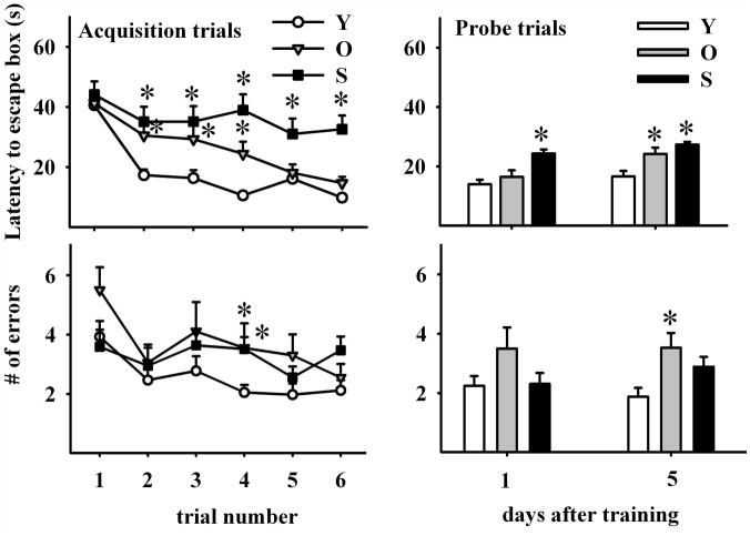
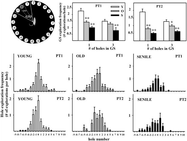
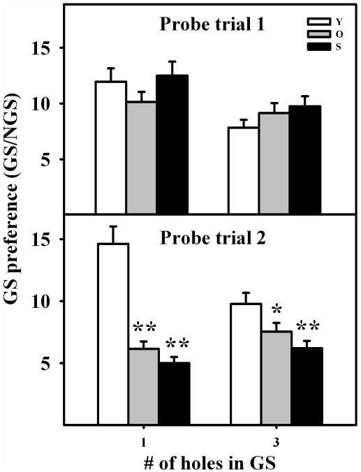
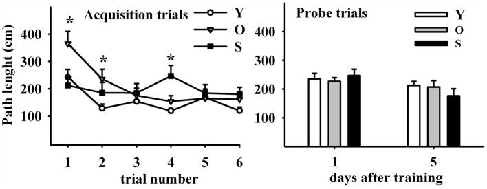
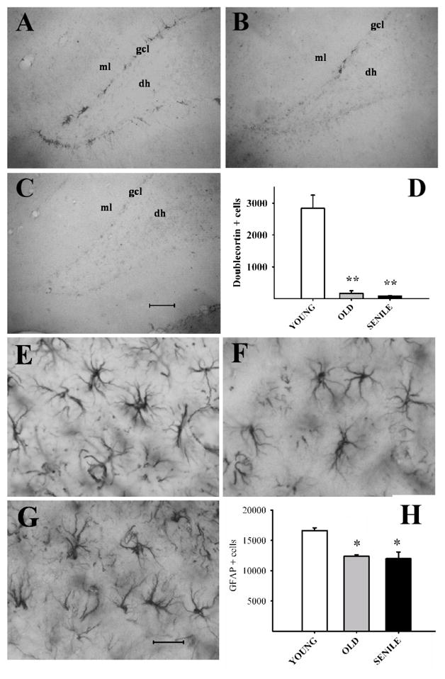
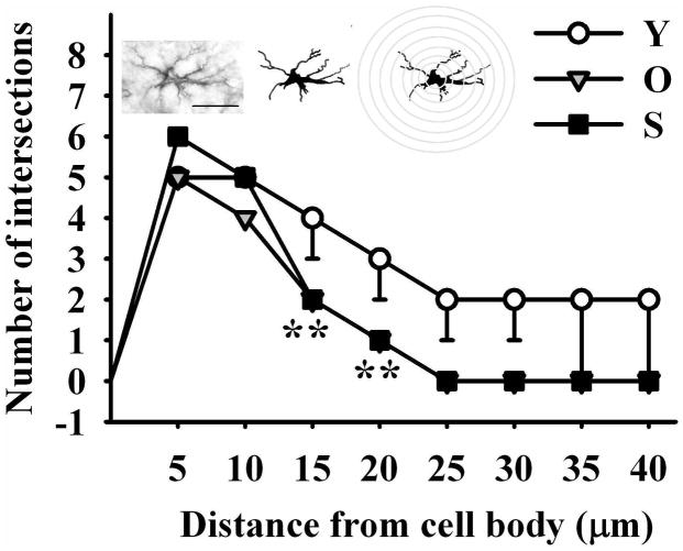
The hippocampus, a medial temporal lobe structure necessary for the formation of spatial memory, is particularly affected by both normal and pathologic aging. In previous studies, we observed a significant age-related increase in dopaminergic neuron loss in the hypothalamus and the substantia nigra of female rats, which becomes more conspicuous at extreme ages. Here, we extend our studies by assessing spatial memory in 4-6 month-old (young), 26-month-old (old) and 29-32-month-old (senile) Sprague-Dawley female rats as well as the age-related histopathological changes in their dorsal hippocampus. Age changes in spatial memory performance were assessed with a modified version of the Barnes maze test. We employed two probe trials (PTs), one and five days after training, respectively, in order to evaluate learning ability as well as short-term and longer-term spatial memory retention. A set of relevant hippocampal cell markers was also quantitated in the animals by means of an unbiased stereological approach. The results revealed that old rats perform better than senile rats in acquisition trials and young rats perform better than both aging groups. However, during short-term PT both aging groups showed a preserved spatial memory while in longer-term PT, spatial memory showed deterioration in both aged groups. Morphological analysis showed a marked decrease (94-97%) in doublecortin neuron number in the dentate gyrus in both aged groups and a reduction in glial fibrillary acidic protein-positive cell number in the stratum radiatum of aging rats. Astroglial process length and branching complexity decreased in aged rats. We conclude that while target-seeking activity and learning ability decrease in aged females, spatial memory only declines in the longer-term tests. The reduction in neuroblast number and astroglial arborescence complexity in the dorsal hippocampus are likely to play a role in the cognitive deficits of aging rats.

Keywords: Barnes maze; GFAP; aging; doublecortin; female-spatial memory; hippocampus.


Figures

Figure 1
Figure 1. Barnes Maze design used in the present study
The maze consists of a black acrylic circular platform, 122 cm in diameter, containing twenty holes around the periphery. The holes are of uniform diameter (10 cm) and appearance, but only one hole is connected to a black escape box. An opaque squared starting chamber is used to place the rats on the platform. Four proximal visual cues are located in the room, 50 cm away from the circular platform. The escape hole is numbered as hole 0 for graphical normalized representation purposes, the remaining holes being numbered 1 to 10 clockwise, and −1 to −9 counterclockwise. Hole 0 remains in a fixed position, relative to the cues in order to avoid randomization of the relative position of the escape box.
Figure 2
Figure 2. Effect of age on learning and spatial memory retention
Upper panels show escape hole latency throughout training (upper left panel) and in probe trials (upper right panel). Lower panels show the number of errors during training (left lower panel) and probe trials (right lower panel). Learning ability was assessed by performing six acquisition trials during three days (2 AT per day). Note that young rats show the greatest overall reduction in primary errors (errors made before the first exploration of hole 0) during acquisition. All data are represented as mean ∀ SEM. Comparisons are made versus the corresponding young data point. *: P<0.05; Number of young, old and senescent rats for AT was 30, 20 and 15 and was 42, 20 and 37 for PT, respectively.
Figure 3
Figure 3. Hole exploration frequency in probe trials
Notice in the middle and lower panels, the bell-shaped distribution of frequencies around hole #0 and the age-related deterioration of this bell shape. Center and right upper panels show goal sector exploration in young, old and senescent rats. Exploratory activity was measured when GS was taken as only hole #0 (delineated by dashed lines) or when the GS consisted of holes −1, 0 and 1 (delineated by solid lines), as illustrated on the left upper panel. Number of young, old and senescent rats for PT1 was 30, 19 and 21 and for PT2 was 29, 20 and 24, respectively. *P<0.05; **P<0.01; Tukey’s post hoc test.
Figure 4
Figure 4. Goal Sector Preference in young, old and senile rats
Columns represent the ratio, GS explorations per hole/NGS explorations per hole, in the different age groups, for GS consisting of either hole# 0 or holes −1,0,1. This ratio provides an index of the accuracy with which rats of the same age group remember a given GS. Notice that this ratio is independent from the target-seeking activity level of rats in a given age group. Statistical comparisons are made versus the young group of each set. *: P<0.05; **: P<0.01. Number of young, old and senescent rats for PT1 was 30, 19 and 21 and for PT2 was 29, 20 and 24, respectively. Other details are as in Fig. 2.
Figure 5
Figure 5. Path length evolution throughout training and probe trials
Left Panel illustrates six acquisition trials during three days (at 2 AT per day). Distance traveled in the maze was progressively reduced in young and old but not in senile rats. Right Panel shows path length in two probe trials performed four days apart, in young, old and senile rats. Path length was no different among age groups. N values and other details are as in Fig 2. The Holm-Sidak post hoc test was used for group comparisons
Figure 6
Figure 6. Mean Velocity evolution throughout training
Left Panel illustrates six acquisition trials during three days. Mean velocity in the maze progressively increased in young but not in old and senile rats. Note that old rats began with higher speeds than their young counterparts. Right Panel shows mean velocity in two probe trials five days apart. Senile rats showed a significantly lower speed than young and old counterparts in both probe trials. (Holm-Sidak pos-hoc method applied to probe trial 1 and Dunn’s Method for probe trial 2). N values and other details are as in Fig 2.
Figure 7
Figure 7. Doublecortin (DCX) and glial fibrillary acidic protein (GFAP) expression in the dorsal hippocampus of young, old and senile rats
Images: Coronal sections of the dentate gyrus in representative animals of each age group showing DCX(+) neurons (Panels A–C) and of the stratum radiatum for GFAP (+) cells (Panels E–G). Scale bars: 100 μm for DCX images and 25 μm for GFAP. DCX and GFAP cell numbers are plotted in Panel D and H, respectively. Note the sharp age related fall in DCX cell numbers. Abbreviations: dh (dentate hilus); gcl (granular cell layer); ml (molecular layer). Number of young, old and senescent hippocampi assessed for DCX was 6 for the 3 age groups and was 5, 6 and 5, respectively for GFAP. Other details are as in Fig. 2. The Holm-Sidak post hoc test was used for group comparisons in both plots.
Figure 8
Figure 8. Impact of age on the length and complexity of glial processes in the stratum radiatum
Astroglial process numbers and length were drawn on a printed image, scaled up and analyzed with NIH software ImageJ running the Sholl Analysis Plugin v1.0. As shown by the scheme over the graph, a grid with concentric rings or shells distributed at equal distances d centered on the soma of a cell was superimposed on GFAP immunoreactive astrocyte images. The number of process intersections i per shell was computed and branching complexity evaluated. The length of the processes was estimated by the sum of the products of d by i for each ring. Holm-Sidak post-hoc test; Double asterisks represent highly significant differences (P<001) of aged animals versus the corresponding young control for the indicated points. The number of hippocampi assessed was 3 for each age group. Scale bar: 20 μm.

References

    1. Azcoitia I1, Perez-Martin M, Salazar V, Castillo C, Ariznavarreta C, Garcia-Segura LM, Tresguerres JA. Growth hormone prevents neuronal loss in the aged rat hippocampus. Neurobiol Aging. 2005;26:697–703. - PubMed
    1. Barnes CA. Memory deficits associated with senescence: a neurophysiological and behavioral study in the rat. J Comp Physiol Psychol. 1979;93:74–104. - PubMed
    1. Barnes CA, Nadel L, Honig WK. Spatial memory deficit in senescent rats. Can J Psychol. 1980;34:29–39. - PubMed
    1. Barrett GL, Bennie A, Trieu J, Ping S, Tsafoulis C. The chronology of ag related spatial learning impairment in two rat strains, as tested by the Barnes maze. Behav Neurosci. 2009;123:533–538. - PubMed
    1. Begega A1, Cuesta M, Rubio S, Méndez M, Santín LJ, Arias JL. Functional Networks involved in spatial learning strategies in middle-aged rats. Neurobiol Learn Mem. 2012;97:346–353. - PubMed

Publication types