. 2015 Jul 3;4(8):980-92.
doi: 10.1242/bio.012773.

Region and dynamic specificities of adult neural stem cells and oligodendrocyte precursors in myelin regeneration in the mouse brain

Affiliations

Region and dynamic specificities of adult neural stem cells and oligodendrocyte precursors in myelin regeneration in the mouse brain

Béatrice Brousse et al. Biol Open. .

Erratum in

Abstract

Myelin regeneration can occur in the brain following demyelination. Parenchymal oligodendrocyte progenitors (pOPC) are known to play a crucial role in this process. Neural stem cells (NSC) residing in the ventricular-subventricular zone (V-SVZ) also have the ability to generate oligodendrocytes but their contribution to endogenous myelin repair was so far considered to be negligible. Here, we addressed the relative contribution of pOPC and V-SVZ-derived neural progenitors (SVZdNP) to remyelination in cuprizone mouse models of acute or chronic corpus callosum (CC) demyelination. Using genetic tracing, we uncover an unexpected massive and precocious recruitment of SVZdNP in the anterior CC after acute demyelination. These cells very quickly adopt an oligodendrocytic fate and robustly generate myelinating cells as efficiently as pOPC do. In more posterior areas of the CC, SVZdNP recruitment is less important whereas pOPC contribute more, underlining a regionalization in the mobilization of these two cell populations. Strikingly, in a chronic model when demyelination insult is sustained in time, SVZdNP minimally contribute to myelin repair, a failure associated with a depletion of NSC and a drastic drop of progenitor cell proliferation in V-SVZ. In this context, pOPC remain reactive, and become the main contributors to myelin regeneration. Altogether our results highlight a region and context-dependent contribution of SVZdNP to myelin repair that can equal pOPC. They also raise the question of a possible exhaustion of V-SVZ proliferation potential in chronic pathologies.

Keywords: Adult neural stem cells; Myelin regeneration; Oligodendrocyte precursors; Subventricular zone.


Conflict of interest statement

Competing interests

The authors declare no competing or financial interests.

Figures

Fig. 1.
Fig. 1.
Experimental design. (A) Model of acute demyelination with short-term (5 weeks) cuprizone administration. Mice were injected for 5 consecutive days with tamoxifen to induce recombination 2 weeks before start of cuprizone treatment. Cuprizone was administered in food during 5 weeks. Mice were sacrificed at different time points during demyelination (W3 and W5) and during remyelination (W5+2off and W5+4off). (B) Model of chronic demyelination with long-term (12 weeks) cuprizone administration. Mice were injected for 5 consecutive days with tamoxifen to induce recombination 2 weeks before start of cuprizone treatment. Cuprizone was administered in food during 12 weeks. Mice were sacrificed at the end of cuprizone treatment (W12) and 4 and 8 weeks after the end of cuprizone treatment (W12+4off and W12+8off). (C) Areas analyzed. The corpus callosum (CC) was analyzed at two distinct locations: at an anterior level (in red, Bregma +0.5 to +1) and at a more posterior area at the level of the fornix (in blue, Bregma −0.3 to −0.8). cx, cortex; v, ventricle; cc, corpus callosum.
Fig. 2.
Fig. 2.
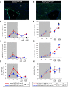
Mobilization of SVZdNP and pOPC in the CC after short-term cuprizone-induced demyelination. (A,B) YFP immunolabeling on coronal sections of the CC of (A) control (CTRL) or (B) cuprizone-treated (W5) NestinCreERT2-YFP mice. While only rare YFP+ cells are observed in the CC of CTL mice, after 5 weeks cuprizone treatment the CC is colonized by numerous YFP+ cells. (C) Quantitative analysis of the YFP+ cell density in the CC at the different time points. (D,E) YFP immunolabeling on coronal sections of (D) the CC of control or (E) cuprizone-treated PDGFRaCre-YFP mice. Note the presence of YFP+ cells in the CC of CTL mice and the presence of cell clusters in mice at the end of the cuprizone treatment. (F) Quantitative analysis YFP+ cell density in the CC at the different time points. (C,F) CUP, cuprizone-treated; CTL, control. Gray block on the graph indicates the period of cuprizone administration. cx, cortex; v, ventricle; cc, corpus callosum. Scale bars 100 µm. Error bars=s.e.m.
Fig. 3.
Fig. 3.
Regionalized mobilization of SVZdNP and pOPC in the CC after short-term cuprizone-induced demyelination. (A) YFP immunolabeling on coronal sections of NestinCreERT2-YFP mice at different time points of cuprizone treatment, focused on the central part of the CC at anterior and posterior levels. Many YFP+ cells are present in the CC. Note the difference between anterior and posterior CC. (B) Quantitative analysis of YFP+ cell density in the CC of NestinCreERT2-YFP mice. (C) YFP immunolabeling on coronal sections of PDGFRaCreERT2-YFP mice at different time points of cuprizone treatment, focused on the central part of the CC at anterior and posterior levels. (D) Quantitative analysis of YFP+ cell density in the CC of PDGFRaCreERT2-YFP mice. (E) Ratio of YFP+ cell density in anterior versus posterior CC in both mouse lines. In NestinCreERT2-YFP mice this ratio is >1 whereas it is <1 in PDGFRaCreERT2-YFP mice showing a regionalization in the contribution of SVZdNP and pOPC to CC repopulation. (B,D,E) CC ant (red), anterior CC; CC post (blue) posterior CC; CTL, control. Gray block on the graph indicates the period of cuprizone administration. Scale bars in A and C, 100 µm. Error bars=s.e.m.; *P<0.05.
Fig. 4.
Fig. 4.
Phenotypic analysis of YFP+ SVZ-derived cells in the CC during acute cuprizone-induced demyelination. (A,B) Triple immunolabeling showing co-expression of YFP, Olig2 and CC1 in numerous cells after 5 weeks cuprizone administration. Arrowheads in B represent YFP+ cells expressing Olig2 but not CC1. Arrows represent YFP+ cells expressing both Olig2 and CC1. (C,D) Quantitative analysis of the proportion of YFP+ cells expressing Olig2, CC1 and GFAP in the (C) anterior CC and (D) posterior CC. (E) Proportion of total CC1+ cells expressing YFP in the CC. CC ant (red), anterior CC; CC post (blue) posterior CC. Gray block on the graph indicates the period of cuprizone administration. (F-H) Myelination potential of SVZdNP is visualized by using mTmG reporter mouse line. Note in F the presence of typical myelin segments. (G,H) immunolabeling showing co-expression of YFP and CC1 (arrowhead in G) and the paranodin labeling concentrate at the tip of GFP+ segments (arrows in H1 and H2). cx, cortex; v, ventricle; cc, corpus callosum. Scale bars: in A, 100 µm; in B, 50 µm; in F, 100 µm; in G, 20 µm and in H, 10 µm. Error bars=s.e.m.
Fig. 5.
Fig. 5.
Phenotypic analysis of pOPC-derived YFP+ cells in the CC during acute cuprizone-induced demyelination. (A) Triple immunolabeling showing that almost all YFP+ cells are Olig2+ (arrows) and only very few cells also express the astrocytic marker GFAP (arrowhead). (B) Double immunolabeling illustrating CC1 expression among YFP+ cells in the CC (arrowhead), arrows show YFP+ cells that do not express CC1. (C,D) Quantitative analysis of the proportion of YFP+ cells expressing Olig2, CC1 and GFAP in the (C) anterior and (D) posterior CC. Scale bars, 50 µm. Error bars=s.e.m.
Fig. 6.
Fig. 6.
Contribution of SVZdNP and pOPC to the oligodendrocytic cell population in the CC during acute cuprizone-induced demyelination. (A) Proportion of Olig2+ cells expressing YFP in the anterior (red) and posterior (blue) CC of both mouse lines (NestinCreERT2-YFP mice in plain lines and PDGFRaCreERT2-YFP mice in dotted lines). (B) YFP+CC1+ cell density in the anterior (ant CC, red) and posterior (post CC, blue) CC of both mouse lines (NestinCreERT2-YFP in plain lines and PDGFRaCreERT2-YFP in dotted lines). Gray blocks on the graphs indicate the period of cuprizone administration. Error bars=s.e.m.
Fig. 7.
Fig. 7.
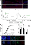
Mobilization of SVZdNP and pOPC in the CC during long-term cuprizone-induced demyelination. (A) YFP immunolabeling on coronal sections of the CC of NestinCreERT2-YFP mice at W12+8. Note the presence of only rare cells in the CC. (B,C) Quantitative analysis of (B) YFP+ and (C) YFP+CC1+ cell density in anterior (ant CC, red) and posterior (post CC, blue) CC of NestinCreERT2-YFP mice. (D) Contribution of SVZdNP to total CC1+ mature oligodendrocyte population in the anterior (red) and posterior (blue) CC. (E) YFP immunolabeling on coronal sections of the CC of PDGFRaCreERT2-YFP mice at W12+8. (F,G) Quantitative analysis of (F) YFP+ and (G) YFP+CC1+ cell density in anterior (red) and posterior (blue) CC of PDGFRaCreERT2-YFP mice. (H) Contribution of pOPC to total CC1+ mature oligodendrocyte population in the anterior (red) and posterior (blue) CC. Gray blocks on the graphs indicate the period of cuprizone administration. cx, cortex; v, ventricle; cc, corpus callosum. Scale bars in A and E, 100 µm. Error bars=s.e.m.
Fig. 8.
Fig. 8.
Effect of long term cuprizone treatment on cell proliferation and oligodendrocyte production in the SVZ. (A,B) PH3 immunolabeling in SVZ in the (A) acute and (B) chronic model at the end point of recovery (W5+4off and W12+8off respectively). Note the drastic reduction of cell proliferation in the lateral wall of the ventricle in the chronic model, even after 8 weeks recovery (B). (C,D) Quantitative analysis of (C) PH3+ and (D) Olig2+ cells in the SVZ of cuprizone-treated (CUP) and control (CTL) mice. Dotted lines symbolize periods without cuprizone (after cuprizone removal; CUP OFF). (E-G) Effect of cuprizone in the neurosphere forming assay. Neurospheres are slightly smaller (E) in presence of 20 µM of cuprizone (Cup) in the culture medium compared to control (DMSO). (G) BrdU and Caspase 3 immunolabeling on neurosphere and quantitative analysis (F) showing that BrdU incorporation is reduced in presence of cuprizone. Note in G the size reduction of neurosphere in presence of cuprizone. Scale bars in A and B, 100 µm; in G, 25 µm. Error bars=s.e.m.; *P<0.05.
Fig. 9.
Fig. 9.
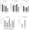
Effect of long-term cuprizone treatment on NSC and neuroblasts. (A) YFP+GFAP+ co-labeling in SVZ shows a decrease in the proportion of NSC among YFP+ cells after 12 weeks but not 5 weeks cuprizone feeding. (B) Neuroblast density in SVZ assessed by doublecortin (DCX) immunolabeling. (C) Neuroblast proliferation in the SVZ showed by the proportion of DCX+ cells in M phase (PH3 labeling). Cuprizone does not affect neuroblast proliferation. (D) Neuroblast density in RMS at the entry of olfactory bulb. Olfactory neurogenesis is transiently affected after 12 weeks cuprizone treatment. (E) Proliferation level in neuroblasts in distal RMS after long-term cuprizone treatment. Note the two-fold increase in the proportion of dividing neuroblasts between W12 and W12+4off. *P<0.05; ns, non significant.

References

    1. Aguirre A., Dupree J. L., Mangin J. M. and Gallo V. (2007). A functional role for EGFR signaling in myelination and remyelination. Nat. Neurosci. 10, 990-1002. 10.1038/nn1938 - DOI - PubMed
    1. Calzolari F., Michel J., Baumgart E. V., Theis F., Götz M. and Ninkovic J. (2015). Fast clonal expansion and limited neural stem cell self-renewal in the adult subependymal zone. Nat. Neurosci. 18, 490-492. 10.1038/nn.3963 - DOI - PubMed
    1. Cayre M., Bancila M., Virard I., Borges A. and Durbec P. (2006). Migrating and myelinating potential of subventricular zone neural progenitor cells in white matter tracts of the adult rodent brain. Mol. Cell. Neurosci. 31, 748-758. 10.1016/j.mcn.2006.01.004 - DOI - PubMed
    1. Curtis M. A., Low V. F. and Faull R. L. M. (2012). Neurogenesis and progenitor cells in the adult human brain: a comparison between hippocampal and subventricular progenitor proliferation. Dev. Neurobiol. 72, 990-1005. 10.1002/dneu.22028 - DOI - PubMed
    1. Cusimano M., Biziato D., Brambilla E., Donega M., Alfaro-Cervello C., Snider S., Salani G., Pucci F., Comi G., Garcia-Verdugo J. M. et al. (2012). Transplanted neural stem/precursor cells instruct phagocytes and reduce secondary tissue damage in the injured spinal cord. Brain 135, 447-460. 10.1093/brain/awr339 - DOI - PMC - PubMed