Review
. 2015 Oct:484:288-304.
doi: 10.1016/j.virol.2015.06.006. Epub 2015 Jul 1.

Recent progress in understanding coxsackievirus replication, dissemination, and pathogenesis

Affiliations
Review

Recent progress in understanding coxsackievirus replication, dissemination, and pathogenesis

Jon Sin et al. Virology. 2015 Oct.

Abstract

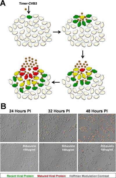
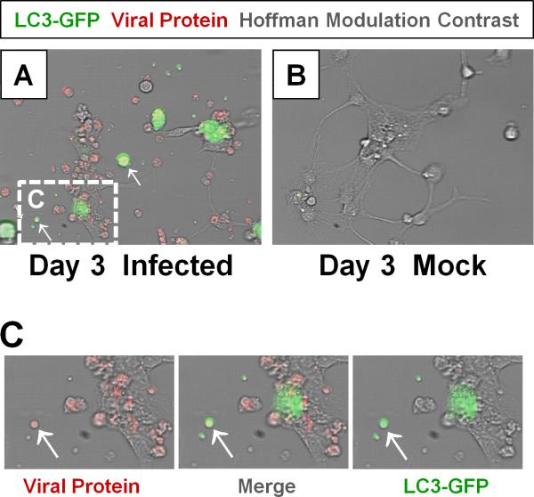
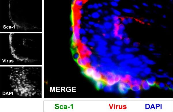
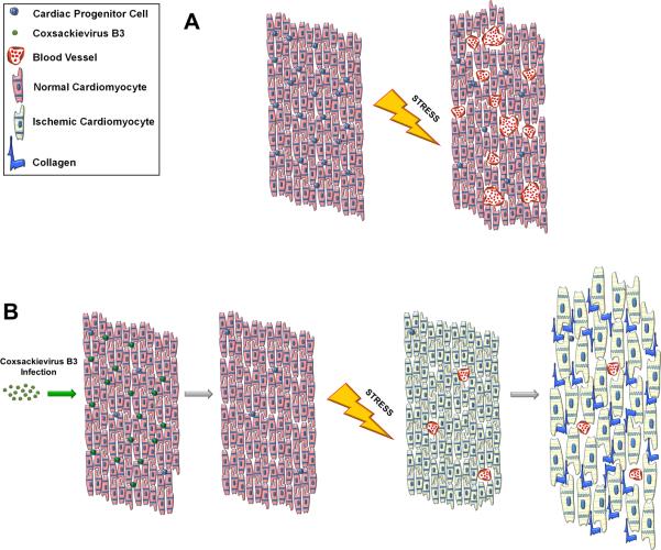
Coxsackieviruses (CVs) are relatively common viruses associated with a number of serious human diseases, including myocarditis and meningo-encephalitis. These viruses are considered cytolytic yet can persist for extended periods of time within certain host tissues requiring evasion from the host immune response and a greatly reduced rate of replication. A member of Picornaviridae family, CVs have been historically considered non-enveloped viruses - although recent evidence suggest that CV and other picornaviruses hijack host membranes and acquire an envelope. Acquisition of an envelope might provide distinct benefits to CV virions, such as resistance to neutralizing antibodies and efficient nonlytic viral spread. CV exhibits a unique tropism for progenitor cells in the host which may help to explain the susceptibility of the young host to infection and the establishment of chronic disease in adults. CVs have also been shown to exploit autophagy to maximize viral replication and assist in unconventional release from target cells. In this article, we review recent progress in clarifying virus replication and dissemination within the host cell, identifying determinants of tropism, and defining strategies utilized by the virus to evade the host immune response. Also, we will highlight unanswered questions and provide future perspectives regarding the potential mechanisms of CV pathogenesis.

Keywords: Autophagy; Cardiac progenitor cells; Coxsackievirus; Enterovirus; Meningoencephalitis; Microvesicles; Myocarditis; Neural progenitor cells; Picornavirus; Virus dissemination.


Figures

Figure 1
Figure 1. HeLa cells infected with Timer-CVB slowly change fluorescence from green to red
The gene for “fluorescent timer” protein was inserted into the infectious plasmid clone for CVB. A) Upon infection with recombinant CVB3 expressing “fluorescent timer” protein (Timer-CVB), the slow conversion of the green fluorescing form of timer protein to red occurred over time in cells overlaid with agar. Initial sites of infection fluoresced red, while newly infected cells fluoresced green. B) HeLa cells infected with Timer-CVB (moi = 0.1) initially fluoresced green (recent viral protein) at 24 hours PI as determined by fluorescence microscopy. By 32 hours PI, both green and red fluorescence (matured viral protein) was observed in infected HeLa cells, and by 48 hours PI the majority of cells fluoresced brightly in the red channel. Fewer green and red infected cells were observed by fluorescence microscopy for HeLa cells treated with the antiviral drug ribavirin (100µg/ml) at every time point.
Figure 2
Figure 2. Detection of LC3 and CVB viral protein in shed EMVs
Differentiated NPSCs transduced with adeno-LC3-GFP were infected with dsRED-CVB (moi = 0.1) and observed by fluorescence microscopy at 3 days PI. (A) Abundant shed EMVs (white arrows) expressing viral protein (red) and a marker for autophagosomes (LC3-GFP, green) were readily observed. (E-F) Higher magnification of (C) showed colocalization of viral protein and LC3-GFP in shed EMVs.
Figure 3
Figure 3. Model of CVB dissemination in the host by shed EMVs
High numbers of LC3II+ extracellular microvesicles (EMVs) containing infectious virus were recently observed following infection of progenitor cells in culture. Both the differentiation process and viral infection may enhance shedding of single membrane EMVs derived from the autophagy pathway. A) Virus-associated EMVs may expand the natural tropism of CV to target cells which fail to express canonical virus receptors. B) Neutralizing antibodies may be ineffective against infectious virus sequestered within the protected environment of the extracellular microvesicle. Also, virus-associated EMVs may increase the stability of infectious virus within the host during hematogenous spread. C) EMVs may assist in viral RNA dissemination during the persistent stage of infection whereby the presence of intact virions and/or structural viral proteins may be limited. D) EMVs may help virions travel and enter new target tissues and cross selectively permeable barriers.
Figure 4
Figure 4. “Bus Stop/Trojan Horse” model for CVB entry across the tight junctions of the blood-CSF barrier
We propose that CVB initially binds to CAR, a tight junction protein, although not entering epithelial cells of the choroid plexus. Upon binding, CCL12 and other chemokines are released by epithelial cells thereby attracting nestin+ myeloid cells which undergo extravasation through tight junctions of choroid plexus epithelial cells. CVB virions enter nestin+ myeloid cells which support infection, and assist with virus entry into the CNS.
Figure 5
Figure 5. CVB productively infected progenitor cells in the juvenile heart
Three day-old mice were infected with eGFP-CVB (105 pfu IP) or mock-infected, and hearts were isolated at 2 days PI. Paraffin-embedded sections of heart tissue were deparaffinized and stained using an antibody against Sca-1 (green) and virus protein (red). Many Sca-1+ cells in heart tissue were shown to be infected with eGFPCVB. DAPI (blue) was utilized to label cell nuclei. Representative images of three infected mice are shown.
Figure 6
Figure 6. Model of adult heart failure in juvenile CVB-infected mice
(A) A population of CPCs susceptible to CVB infection resides within the myocardium. Upon augmented cardiac stress, oxygen demand increases within the heart tissue. CPCs are recruited to drive angiogenesis and neovascularization which increases vascular density in the muscle allowing for efficient perfusion of oxygenated blood. (B) When the heart undergoes mild CVB infection, CPCs are preferentially targeted by the virus resulting in a depletion of the CPC population; however the myocardium is otherwise normal. Following cardiac stress, the limited number of remaining CPCs cannot sufficiently stimulate blood vessel formation and the myocardium becomes ischemic. The lack of vascularization causes the heart to become hypertrophic resulting in scar formation and cardiac dysfunction.

References

    1. Abzug MJ. Presentation, diagnosis, and management of enterovirus infections in neonates. Paediatr.Drugs. 2004;6:1–10. - PubMed
    1. Ahn J, Jee Y, Seo I, Yoon SY, Kim D, Kim YK, Lee H. Primary neurons become less susceptible to coxsackievirus B5 following maturation: the correlation with the decreased level of CAR expression on cell surface. J.Med.Virol. 2008;80(3):434–440. - PubMed
    1. Alderman CP, Moritz CK, Ben-Tovim DI. Abnormal platelet aggregation associated with fluoxetine therapy. Ann.Pharmacother. 1992;26:1517–1519. - PubMed
    1. Alidjinou EK, Sane F, Bertin A, Caloone D, Hober D. Persistent infection of human pancreatic cells with Coxsackievirus B4 is cured by fluoxetine. Antiviral Res. 2015:10. - PubMed
    1. Alirezaei M, Flynn CT, Whitton JL. Interactions between enteroviruses and autophagy in vivo. Autophagy. 2012a;8:973–975. - PMC - PubMed

Publication types

LinkOut - more resources