. 2015 Jul 6:6:7538.
doi: 10.1038/ncomms8538.

Suppression of the alternative lengthening of telomere pathway by the chromatin remodelling factor ATRX

Affiliations

Suppression of the alternative lengthening of telomere pathway by the chromatin remodelling factor ATRX

David Clynes et al. Nat Commun. .

Abstract

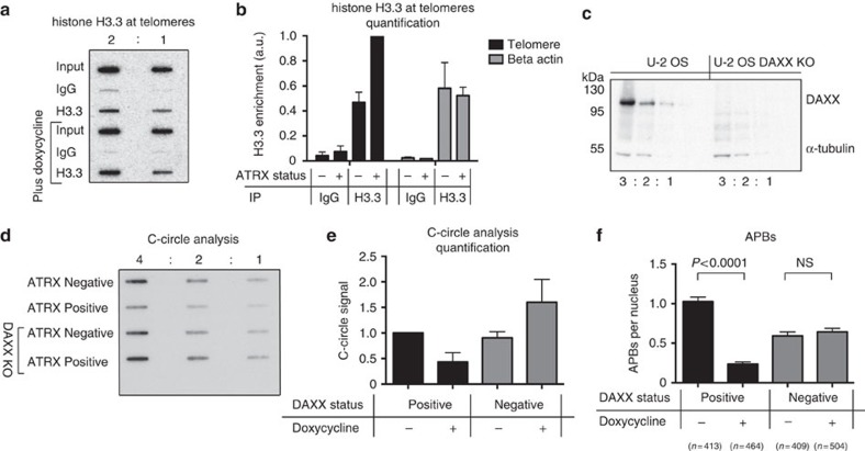
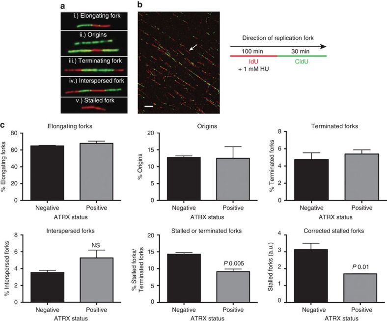
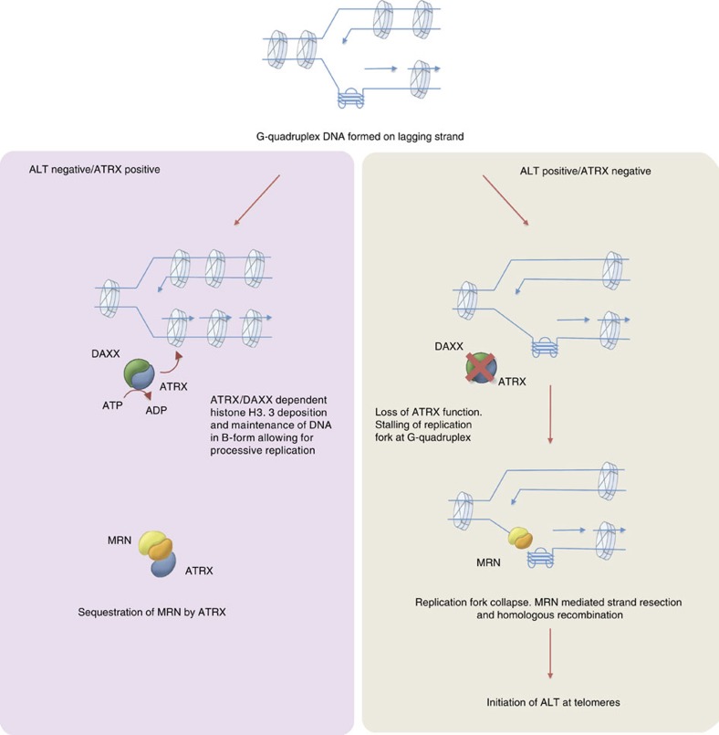
Fifteen per cent of cancers maintain telomere length independently of telomerase by the homologous recombination (HR)-associated alternative lengthening of telomeres (ALT) pathway. A unifying feature of these tumours are mutations in ATRX. Here we show that expression of ectopic ATRX triggers a suppression of the pathway and telomere shortening. Importantly ATRX-mediated ALT suppression is dependent on the histone chaperone DAXX. Re-expression of ATRX is associated with a reduction in replication fork stalling, a known trigger for HR and loss of MRN from telomeres. A G-quadruplex stabilizer partially reverses the effect of ATRX, inferring ATRX may normally facilitate replication through these sequences that, if they persist, promote ALT. We propose that defective telomere chromatinization through loss of ATRX promotes the persistence of aberrant DNA secondary structures, which in turn present a barrier to DNA replication, leading to replication fork stalling, collapse, HR and subsequent recombination-mediated telomere synthesis in ALT cancers.


Figures

Figure 1
Figure 1. Exogenously expressed ATRX represses the ALT pathway.
(a) Immunoblot showing expression of ATRX in U-2 OSATRX cell line on addition of 0.4 μg ml−1 doxycycline for the specified number of days. Alpha tubulin is shown as a loading control. (b) Chromatin Immunoprecipitation showing that on addition of 0.4 μg ml−1 doxycycline for 4 days the exogenously expressed ATRX is targeted to telomeres. Immunoprecipitated DNA was slot blotted and probed with a 32P-labelled TTAGGG probe. (c) Induction of ATRX by doxycycline leads to a fall in C-circles which is reversed on withdrawal of doxycycline at day 5 (shown as a dashed line). (d) Representative immunofluorescence images showing the presence of ALT-associated PML bodies (APBs) in untreated U-2 OSATRX cells and the reduction in APBs on induction of ATRX expression by addition of 0.4 μg ml−1 doxycycline for 4 days. A 10-μm scale marker is shown. APBs (e) and TIFs (f) are scored as a direct co-localization between either TRF2 and PML for APBs, or between TRF2 and 53BP1 for TIFs, with the chart showing the average number of APBs or TIFs in U-2 OSATRX cells with and without ATRX expression. Co-localizing foci were scored using the JACoP plugin for ImageJ. Number of cells scored (n) is shown in parentheses. Statistical significance was determined using a Mann–Whitney test. (g) Terminal restriction fragment length analysis using a TTAGGG probe showing progressive telomere shortening on expression of ATRX. (h) Representative image of chromosome orientation fluoresence in situ hybridization (CO-FISH). G-rich telomeric DNA is stained in red. A crossover event is depicted with a white arrow, a 10-μm scale marker is shown. (i) Quantitation of CO-FISH in U-2 OSATRXcells before and after ATRX induction for 4 days. Telomere sister chromatid exchange (TSCE) was scored as a percentage of exchanges per metaphase spread. Error bars denote s.e.m. Over 2,000 telomere ends were scored for cross-overs per treatment group. Statistical significance was determined using a Mann–Whitney test.
Figure 2
Figure 2. Re-introduction of ATRX increases levels of telomeric histone H3.3.
(a) Representative blot showing H3.3 ChIP in the absence or presence of ATRX (following 4 days treatment with 0.4 μg ml−1 doxycycline). Immunoprecipitated DNA was slot blotted and probed with a TTAGGG probe. (b) Graph showing average quantitation of H3.3 ChIP assays from three biological replicates. Levels of H3.3 at beta-actin were assessed by qPCR and did not change on re-expression of ATRX. Error bars indicate ±s.e.m. Enrichment is shown relative to ATRX-positive telomeric histone H3.3 levels. (c) Immunoblot showing serial dilutions of a U-2 OSATRX clone in which DAXX was successfully knocked out. Alpha tubulin is shown as a loading control. (d) Slot blot of a representative C-circle assay in U-2 OSATRX and DAXX null clone in the absence or presence of ATRX (induced for 4 days). Serial dilutions are shown. Quantification of three biological replicates is shown in (e). (f) Quantification of three biological replicates of APB immunofluorescence in U-2 OSATRXand DAXX null cells before and after ATRX induction for 4 days. Co-localizing foci were analysed as indicated in Fig. 1d. Statistical significance was determined using a Mann–Whitney test.
Figure 3
Figure 3. Treatment with a G4-stabilizing ligand impedes ability of ATRX to suppress ALT.
Cells were treated for 4 days with 0.4 μg ml−1 doxycycline and PDS treatment after day 2 for 2 days (a) Representative immunofluoresence images showing output of co-localization analysis between PML and TRF2 as displayed using HTML5 PIVOT software. Row 3 shows foci (displayed in white) called by ImageJ and row 4 shows the number and position of co-localizing foci, representing APBs. A 10-μm scale marker is shown. (b) Quantitation of APB numbers per nuclei, number of cells scored is shown in parentheses. Statistical significance was determined using a Mann–Whitney test. (c) Graph displaying average quantitation of three C-circle assay biological replicates, showing a higher level of C-circles on treatment with PDS in cells treated with 0.4 μg ml−1 doxycycline for 4 days. Statistical significance was determined using a Mann–Whitney test. Original representative blot is shown in Supplementary Fig. 4c.
Figure 4
Figure 4. Exogenously expressed ATRX leads to a reduction in replication fork stalling.
(a) Representative images of five classes of replication intermediates identified in this study. (b) Representative image of actual fibres from U-2 OSATRX cells with a white arrow indicating a stalled fork, a 10 μm scale marker is shown. (c) Relative frequency of replication intermediates in ATRX-negative and ATRX-positive (4 days treatment with 0.4 μg/ml doxycycline) U-2 OSATRX cells. Over 1,400 fibres totalled from three independent replicates were scored and error bars indicate±s.e.m. Statistical significance was determined using a Mann–Whitney test. Stalled forks were calculated as a ratio of the % stalled or terminated forks and terminated forks.
Figure 5
Figure 5. Exogenously expressed ATRX in U-2 OSATRX cells interacts with the MRN complex and alters its localization.
(a) Nuclear extracts from U-2 OSATRX cell line with and without exogenously expressed ATRX were immunoprecipitated with both RAD50 and MRE11 antibodies. For no IP beads blocked beads were added without prior antibody coupling. The presence of ATRX in immunoisolated interaction partners was assessed by western blotting. (b) Immunofluorescence with white arrows showing co-localization between exogenously expressed ATRX and MRE11 in the U-2 OSATRX cell line in two representative nuclei. A 10-μm scale marker is shown. (c) Representative blot showing MRE11 ChIP in both the absence and presence of ATRX (following 4 days treatment with 0.4 μg ml−1 doxycycline). Immunoprecipitated DNA was slot blotted and probed with a 32P-labelled TTAGGG probe. (d) Graph showing average quantitation of MRE11 at telomeres by ChIP assay from three biological replicate. Error bars indicate ±s.e.m. (e,f) Immunofluorescence showing loss in co-localization (white arrows) between MRE11 and telomeric marker TRF2 or (g,h) MRE11 and PML on expression of ATRX (13 days treatment with 0.4 μg ml−1 doxycycline). Number of cells scored are shown in parentheses. Statistical significance was determined using a Mann–Whitney test. A 10-μm scale marker is shown.
Figure 6
Figure 6. Model for ATRX-mediated suppression of the ALT pathway.
ATRX together with DAXX deposits histone H3.3 at telomeres, which in turn may facilitate DNA replication through G-quadruplex sequences. The presence of G-quadruplex structures in an ATRX null tumour cell leads to replication fork stalling and collapse, providing a substrate for MRN-dependent homologous recombination and maintenance of telomere length through ALT. ATRX additionally interacts with the MRN complex, facilitating its distribution away from PML bodies and telomeres, further limiting HR.

References

    1. Gilson E. & Geli V. How telomeres are replicated. Nat. Rev. Mol. Cell Biol. 8, 825–838 (2007). - PubMed
    1. Bryan T. M., Englezou A., Dalla-Pozza L., Dunham M. A. & Reddel R. R. Evidence for an alternative mechanism for maintaining telomere length in human tumors and tumor-derived cell lines. Nat. Med. 3, 1271–1274 (1997). - PubMed
    1. Dunham M. A., Neumann A. A., Fasching C. L. & Reddel R. R. Telomere maintenance by recombination in human cells. Nat. Genet. 26, 447–450 (2000). - PubMed
    1. Perrem K. et al.. Repression of an alternative mechanism for lengthening of telomeres in somatic cell hybrids. Oncogene 18, 3383–3390 (1999). - PubMed
    1. Jiao Y. et al.. DAXX/ATRX, MEN1, and mTOR pathway genes are frequently altered in pancreatic neuroendocrine tumors. Science 331, 1199–1203 (2011). - PMC - PubMed

Publication types