

Electrochemical characterization of MC3T3-E1 cells cultured on γTiAl and Ti-6Al-4V alloys
- PMID: 26145813
- PMCID: PMC4565729
- DOI: 10.1016/j.bioelechem.2015.06.012
Electrochemical characterization of MC3T3-E1 cells cultured on γTiAl and Ti-6Al-4V alloys
Abstract
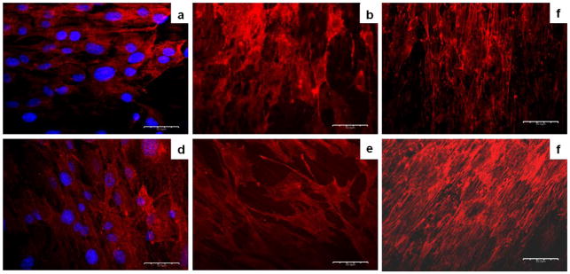
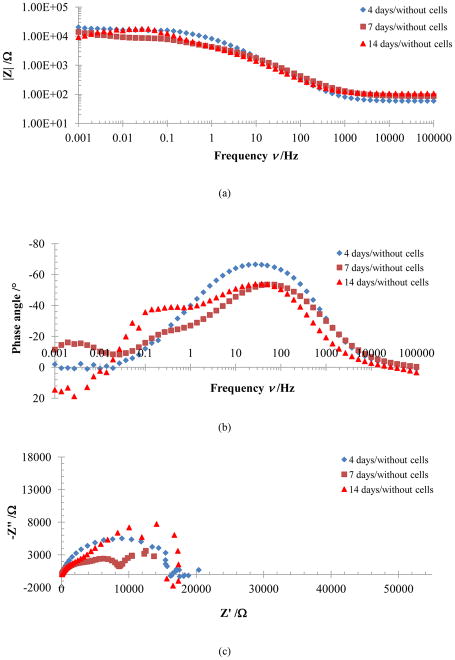
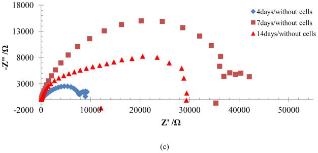
Electrochemical impedance spectroscopy (EIS) was used to study the behavior of MC3T3-E1 cells cultured in an αMEM+FBS solution on two Ti-based alloys (Ti-6Al-4V and γTiAl) for 4, 7 and 14 days. EIS measurements were carried out at an open-circuit potential in a 1 mHz to 100 kHz frequency range. Results indicate a general increase in impedance on the Ti alloy surfaces with cells as a function of time. Bode plots indicate changes corresponding to the passive oxide film, adsorption of proteins and cell tissue on surfaces with the passage of time. Normal cellular activity based on the polygonal morphology, with long and fine cytoplasmic prolongations of the cells on Ti-6Al-4V and γTiAl was observed from SEM images. Similarly, mineralization nodules corresponding to cell differentiation associated with the osseogenetic process were observed confirmed by Alizarin Red S staining. Immunofluorescence analysis to detect the presence of collagen Type I showed an increase in the segregation of collagen as a function of time. The impedance values obtained from EIS testing are indicative of the corrosion protection offered to the Ti alloy substrates by the cell layer. This study shows that γTiAl has better corrosion resistance than that of Ti-6Al-4V in the αMEM+FBS environment in the presence of MC3T3-E1 cells.
Keywords: EIS; Electrochemical characterization; MC3T3-E1 cells; Ti alloys.
Copyright © 2015 Elsevier B.V. All rights reserved.
Conflict of interest statement
We have no conflict of interest based on this work with any person or entity.
Figures


References
-
- Bhat SV. Biomaterials. 2. Alpha Science; Middlesex, UK: 2005.
-
- Hanawa T. Evaluation techniques of metallic biomaterials in vitro. Sci Technol Adv Mater. 2002;3:289–295.
-
- Hiromoto S, Noda K, Hanawa T. Development of electrolytic cell with cell-culture for metallic biomaterials. Corros Sci. 2002;44:955–965.
-
- Huang HH. In situ surface electrochemical characterizations of Ti and Ti-6Al-4V alloy cultured with osteoblast-like cells. Biochem Biophys Res Co. 2004;314:787–792. - PubMed
-
- García-Alonso MC, Saldaña L, Vallés G, González-Carrasco JL, González-Cabrero J, Martínez ME, Gil-Garay E, Munuera L. In vitro corrosion behaviour and osteoblast response of thermally oxidised Ti6Al4V alloy. Biomaterials. 2003;24:19–26. - PubMed
Publication types
MeSH terms
Substances
Grants and funding
LinkOut - more resources
Full Text Sources
Other Literature Sources
