. 2015 Jul 4:5:36.
doi: 10.1186/s13578-015-0026-x. eCollection 2015.

Hypoxia induces calpain activity and degrades SMAD2 to attenuate TGFβ signaling in macrophages

Affiliations

Hypoxia induces calpain activity and degrades SMAD2 to attenuate TGFβ signaling in macrophages

Wei Cui et al. Cell Biosci. .

Abstract

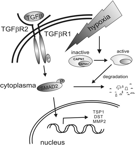
Background: Under inflammatory conditions or during tumor progression macrophages acquire distinct phenotypes, with factors of the microenvironment such as hypoxia and transforming growth factor β (TGFβ) shaping their functional plasticity. TGFβ is among the factors causing alternative macrophage activation, which contributes to tissue regeneration and thus, resolution of inflammation but may also provoke tumor progression. However, the signal crosstalk between TGFβ and hypoxia is ill defined.

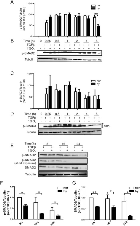
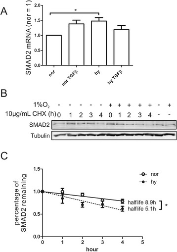
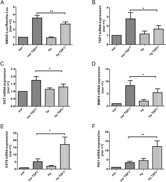
Results: Exposing human primary macrophages to TGFβ elicited a rapid SMAD2/SMAD3 phosphorylation. This early TGFβ-signaling remained unaffected by hypoxia. However, with prolonged exposure periods to TGFβ/hypoxia the expression of SMAD2 declined because of decreased protein stability. In parallel, hypoxia increased mRNA and protein amount of the calpain regulatory subunit, with the further notion that TGFβ/hypoxia elicited calpain activation. The dual specific proteasome/calpain inhibitor MG132 and the specific calpain inhibitor 1 rescued SMAD2 degradation, substantiating the ability of calpain to degrade SMAD2. Decreased SMAD2 expression reduced TGFβ transcriptional activity of its target genes thrombospondin 1, dystonin, and matrix metalloproteinase 2.

Conclusions: Hypoxia interferes with TGFβ signaling in macrophages by calpain-mediated proteolysis of the central signaling component SMAD2.

Keywords: Calpain; Hypoxia; Macrophages; SMAD2 degradation; TGFß; phospho-SMAD2.


Figures

Fig. 1
Fig. 1
Hypoxia attenuates TGFß-induced SMAD2 phosphorylation in macrophages. Time-dependent Western blot analysis b, d and the corresponding statistical evaluation a, c of SMAD2 a, b and SMAD3 c, d phosphorylation in human primary macrophages exposed to TGFß under normoxia (−; nor) or hypoxia (1 % O2; hy). Macrophages were exposed to TGFß under normoxia vs. hypoxia for 8, 16, and 24 h, followed by (e) Western blot analysis of total SMAD2 and SMAD2 phosphorylation and corresponding statistical analysis f, g. Tubulin served as a loading control
Fig. 2
Fig. 2
Expression of TGFBR2 and SMAD7 in response to TGFß/hypoxia. Macrophages were transfected with non-targeting siRNA constructs (ctr), siRNA-HIF1α (si1α), or siRNA-HIF2α (si2α) and mRNA expression of TGFBR2 a or SMAD7 b were analyzed by quantitative PCR after treatments with TGFß under normoxia (nor) or hypoxia (hy) for 8 h. c Cell surface expression of TGFBR2 was followed by flow cytometry after stimulation of macrophages with TGFß under normoxia (nor) vs. hypoxia (hy) for 8 h and d mean fluorescence intensity was calculated from 4 individual experiments. e, f Time-dependent Western blot analysis and statistical evaluation of SMAD7 expression in macrophages exposed to TGFß under normoxia or hypoxia. g, h Macrophages were exposed to the phosphatase inhibitor okadaic acid (OA) 1 h prior to adding TGFß for 8 h. Phosphorylation and total protein expression of SMAD2 was followed by Western blot analysis, using tubulin as a loading control
Fig. 3
Fig. 3
SMAD2 protein stability is reduced in response to TGFß/hypoxia. a mRNA expression of SMAD2 followed by quantitative PCR in macrophages stimulated for 8 h with TGFß under normoxia (nor) vs. hypoxia (hy). b Macrophages were stimulated with TGFß for 4 h, followed by the addition of cycloheximide for 0 h to 4 h and subsequent Western blot analysis of SMAD2 expression. c Half-life determination of SMAD2 in TGFß-stimulated macrophages under normoxia (nor) vs. hypoxia (hy) as a result of experiments performed in Fig. 3b
Fig. 4
Fig. 4
SMAD2 degradation under hypoxia is facilitated by calpain. a-f Macrophages were stimulated with TGFß under normoxia vs. hypoxia for 8 h in the presence of bafilomycin a, b, MG132 c, d, or lactacystin e, f. 2 h after TGFß, inhibitors were added and incubations went on for 6 h. Western blot analysis and statistical evaluation is presented. g mRNA expression of the calpain regulatory subunit (CAPNS1) was analyzed by quantitative PCR in macrophages exposed for 8 h to TGFß under normoxia (nor) vs. hypoxia (hy). h Western blot analysis of the calpain regulatory subunit after exposure to TGFß and/or hypoxia for 8 h. i mRNA expression of the calpain 1 catalytic subunit (CAPN1) was analyzed by quantitative PCR in macrophages exposed for 8 h to TGFß under normoxia (nor) vs. hypoxia (hy). j Proteolytic cleavage of CAPN1 under TGFß/hypoxia was followed in macrophages by Western blot analysis after 8 h. k Macrophages were exposed to the calpain inhibitor 1 (CPI) to follow SMAD2 degradation after stimulating cells for 8 h with TGFß under normoxia vs. hypoxia
Fig. 5
Fig. 5
Hypoxia reduces SMAD2-dependent transcription. a TGFß-induced transcriptional activity in J774A.1 cells transfected with a SBE4-Luc (4xSMAD binding element coupled to luciferase) as well as a Renilla-Luc plasmid. Stimulation was with TGFß under normoxia (nor) vs. hypoxia (hy) for 8 h. mRNA expression of b thrombospondin1 (TSP1), c dystonin DST, d matrix metalloproteinase (MMP2), e cystatin (CST6), and (f) plasminogen activator inhibitor 1 (PAI1) was analyzed by quantitative PCR in macrophages exposed for 8 h to TGFß under normoxia (nor) vs. hypoxia (hy)
Fig. 6
Fig. 6
Graphical abstract. TGFβ binds to its receptor (TGFβR1 and TGFβR2), induces phosphorylation of TGFβR1 and downstream signaling molecules like SMAD2. Active SMAD2 translocates to the nucleus to provoke expression of target genes, i.e., TSP1, DST, and MMP2. Hypoxia in combination with TGFβ activates calpain to degrade SMAD2, which in turn attenuates SMAD2-dependent transcriptional activation and thus, diminishes the induction of TSP1, DST, and MMP2

References

    1. Murray PJ, Allen JE, Biswas SK, Fisher EA, Gilroy DW, Goerdt S, et al. Macrophage activation and polarization: nomenclature and experimental guidelines. Immunity. 2014;41(1):14–20. doi: 10.1016/j.immuni.2014.06.008. - DOI - PMC - PubMed
    1. Weigert A, Brüne B. Nitric oxide, apoptosis and macrophage polarization during tumor progression. Nitric Oxide. 2008;19(2):95–102. doi: 10.1016/j.niox.2008.04.021. - DOI - PubMed
    1. Shi Y, Massagué J. Mechanisms of TGF-β signaling from cell membrane to the nucleus. Cell. 2003;113(6):685–700. doi: 10.1016/S0092-8674(03)00432-X. - DOI - PubMed
    1. Derynck R, Zhang YE. Smad-dependent and Smad-independent pathways in TGF-β family signalling. Nature. 2003;425(6958):577–84. doi: 10.1038/nature02006. - DOI - PubMed
    1. Nurgazieva D, Mickley A, Moganti K, Ming W, Ovsyi I, Popova A, et al. TGF-β1, but not bone morphogenetic proteins, activates Smad1/5 pathway in primary human macrophages and induces expression of proatherogenic genes. J Immunol. 2015;194(2):709–18. doi: 10.4049/jimmunol.1300272. - DOI - PubMed

LinkOut - more resources