. 2015 Jul;3(7):894-907.
doi: 10.1039/C5BM00006H.

Shape Control in Engineering of Polymeric Nanoparticles for Therapeutic Delivery

Affiliations

Shape Control in Engineering of Polymeric Nanoparticles for Therapeutic Delivery

John-Michael Williford et al. Biomater Sci. 2015 Jul.

Abstract

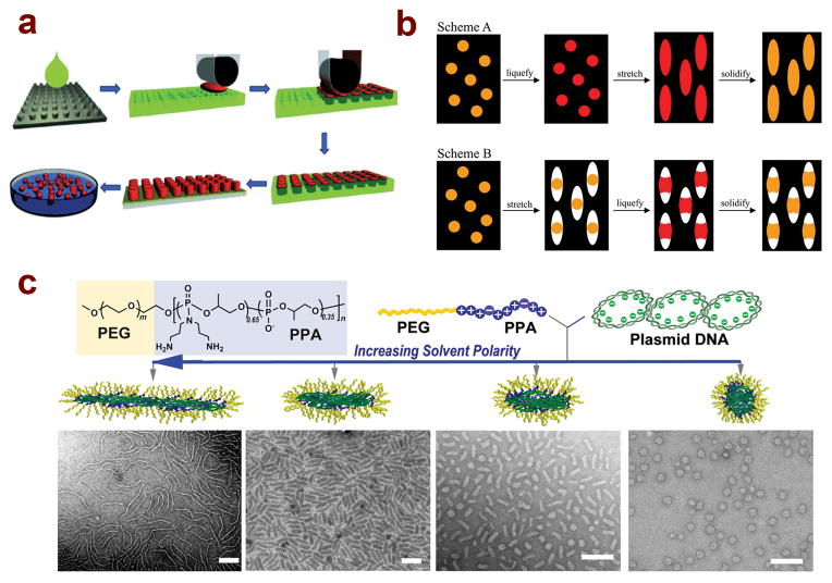
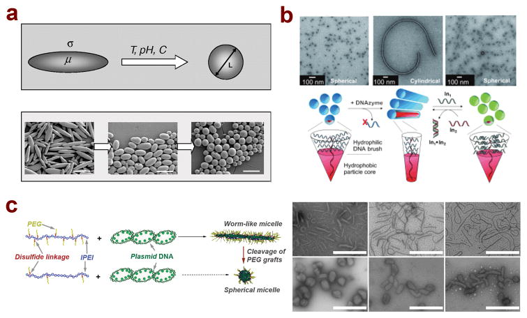
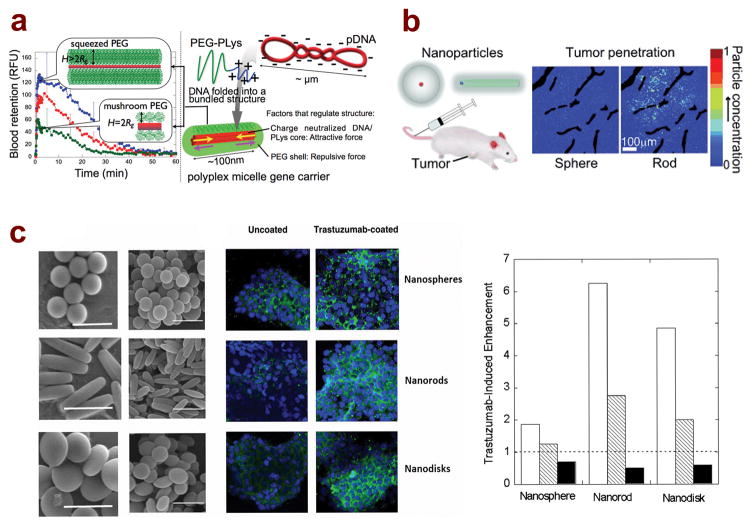
Nanoparticle-mediated delivery of therapeutics holds great potential for the diagnosis and treatment of a wide range of diseases. Significant advances have been made in the design of new polymeric nanoparticle carriers through modulation of their physical and chemical structures and biophysical properties. Nanoparticle shape has been increasingly proposed as an important attribute dictating their transport properties in biological milieu. In this review, we highlight three major methods for preparing polymeric nanoparticles that allow for exquisite control of particle shape. Special attention is given to various approaches to controlling nanoparticle shape by tuning copolymer structural parameters and assembly conditions. This review also provides comparisons of these methods in terms of their unique capabilities, materials choices, and specific delivery cargos, and summarizes the biological effects of nanoparticle shape on transport properties at the tissue and cellular levels.

Keywords: drug delivery; gene therapy; polymeric nanoparticles; shape control.


Figures

Figure 1
Figure 1
Several methods have been developed to control the shape of polymeric nanoparticles for therapeutic delivery applications. (A) PRINT technology allows for the generation of particles with controlled shapes and surface chemistries through harvesting from polymer molds with low surface energy; (reprinted with permission, ©2013 Wiley-VCH Verlag GmbH & Co. KGaA, Weinheim) (B) Two distinct methods for generating nanoparticles with nonspherical shapes through stretching and liquefication of precursor films, leading to the formation of rod shaped and barrel shaped particles; (©2007 National Academy of Sciences, USA) (C) Self-assembly of PEG-polycation/DNA nanoparticles in solutions with varying solvent polarity leads to the formation of different shapes, including spheres, rod-like, and worm-like particles. (reprinted with permission, ©2013 Wiley-VCH Verlag GmbH & Co. KGaA, Weinheim.)
Figure 2
Figure 2
Several recent studies have developed polymeric nanoparticles capable of shape transformation in response to external stimuli. (A) PLGA polymer particles can transform their shape from rod-like to spherical on time scales ranging from minutes to hours in response to external triggers such as various chemicals, pH changes, and temperature changes; (B) Self-assembled DNA brush polymer micelles undergo shape transformation from spherical to cylindrical particles upon enzymatic cleavage of a fraction of the brush segment, which can be reversed by re-introducing a similar DNA segment through complementary base pairing; (reprinted with permission, ©2010 Wiley-VCH Verlag GmbH & Co. KGaA, Weinheim) (C) Shape transformation of polymer/DNA micelles can be achieved through cleavage of a fraction of the PEG chains on the micelle surface, leading to a transition from worm-like shapes to more condensed spherical and short rod shapes. (Reproduced by permission of The Royal Society of Chemistry.)
Figure 3
Figure 3
Nanoparticle shape influences the biological response both in vitro and in vivo for drug and gene delivery applications. (A) Nanoparticle shape is influenced by the crowdedness of the PEG layer on the surface of polymer/DNA micelles. Dense PEG layer leads to the formation of longer rod shapes that, upon systemic administration via tail vein injection, leads to extended circulation compared to shorter, rod shaped micelles; (reprinted with permission, ©2013 American Chemical Society) (B) Greater tumor penetration is observed following i.v. injection of spherical and rod-shaped silica-quantum dot nanoparticles; (reprinted with permission, ©2011 Wiley-VCH Verlag GmbH & Co. KGaA, Weinheim) (C) Scanning electron microscopy images of polystyrene nanospheres, nanorods, and nanodisks, as well as fluorescent microscopy images of in vitro cellular uptake of shaped nanoparticles (green) in BT-474 breast cancer cells (blue) comparing uncoated and antibody (trastuzumab)-coated particles. Graph shows that trastuzumab coating enhancement is greatest for nanorods, followed by nanodisks and nanospheres in both BT-474 cells (white bars) and SK-BR-3 cells (dashed bars). No enhancement was observed for any shape in MDA-MB-231 cells (black bars).

References

    1. Davis ME, Chen Z, Shin DM. Nat Rev Drug Discov. 2008;7:771–782. - PubMed
    1. Parveen S, Misra R, Sahoo SK. Nanomedicine. 2012;8:147–166. - PubMed
    1. Shi JJ, Votruba AR, Farokhzad OC, Langer R. Nano Lett. 2010;10:3223–3230. - PMC - PubMed
    1. Perfezou M, Turner A, Merkoci A. Chem Soc Rev. 2012;41:2606–2622. - PubMed
    1. Pack DW, Hoffman AS, Pun S, Stayton PS. Nat Rev Drug Discov. 2005;4:581–593. - PubMed

Publication types