. 2015:2015:609637.
doi: 10.1155/2015/609637. Epub 2015 Jun 4.

Primary Bronchopulmonary Actinomycosis Masquerading as Lung Cancer: Apropos of Two Cases and Literature Review

Affiliations

Primary Bronchopulmonary Actinomycosis Masquerading as Lung Cancer: Apropos of Two Cases and Literature Review

Stamatis Katsenos et al. Case Rep Infect Dis. 2015.

Abstract

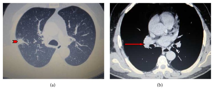
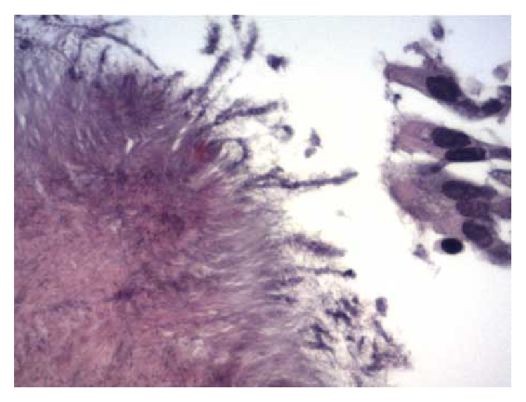
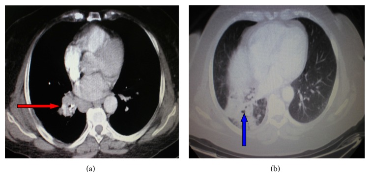
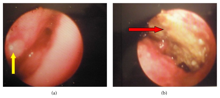
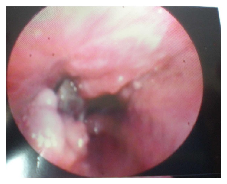
Actinomycosis is a rare and slowly progressive infectious disease that can affect a variety of organ systems including the lung. It is caused by filamentous Gram-positive anaerobic bacteria of the genus Actinomyces. Despite its rarity, pulmonary actinomycosis can involve lung parenchyma, bronchial structures, and chest wall. The disease can mimic lung malignancy given its nonspecific clinical and radiological presentation, thus posing a diagnostic dilemma to the attending physician. In this paper, we describe two patients with pulmonary actinomycosis mimicking bronchogenic carcinoma; the former presented with peripheral infiltrate and associated hilar/mediastinal lymphadenopathy and the latter presented with a foreign body-induced endobronchial mass. Clinical, imaging, diagnostic, and therapeutical aspects of the disease are discussed, demonstrating the paramount importance of the histological examination of lung tissue specimens in the confirmation of the infection given either its low culture yield or the limited use of new molecular diagnostic tools in routine clinical practice.


Figures

Figure 1
Figure 1
(a and b) Contrast-enhanced chest computed tomography showing a nodular opacity in the posterior segment of the right upper lobe (arrowhead) accompanied by mild ipsilateral pleural thickening and bilateral mediastinal lymphadenopathy (red arrow).
Figure 2
Figure 2
Bronchoscopic biopsy specimen demonstrating colonies of organisms with radiating eosinophilic terminal clubs on staining with haematoxylin-eosin (original magnification ×400).
Figure 3
Figure 3
(a and b) A contrast-enhanced computed tomography of the thorax revealing a right hilar mass (red arrow) compressing the bronchus intermedius with accompanying dense airspace opacification of right lower lobe (blue arrow) and atelectasis.
Figure 4
Figure 4
(a and b) Bronchoscopy showing a soft granulation tissue mass (yellow arrow) and an impacted foreign body (red arrow) occluding completely the right bronchus intermedius.
Figure 5
Figure 5
Repeat bronchoscopy after three weeks of antibiotic treatment showing remarkable resolution of the inflammatory mass, thus facilitating foreign body extraction.

References

    1. Wong V. K., Turmezei T. D., Weston V. C. Actinomycosis. BMJ. 2011;343 doi: 10.1136/bmj.d6099.d6099 - DOI - PubMed
    1. Mabeza G. F., Macfarlane J. Pulmonary actinomycosis. European Respiratory Journal. 2003;21(3):545–551. doi: 10.1183/09031936.03.00089103. - DOI - PubMed
    1. Kolditz M., End A. Actinomycosis of the lung and pleura. In: Rohde G., Subotic D., editors. Complex Pleuropulmonary Infections. Vol. 61. 2013. pp. 66–80. (European Respiratory Monograph).
    1. Hall V. Actinomyces-Gathering evidence of human colonization and infection. Anaerobe. 2008;14(1):1–7. doi: 10.1016/j.anaerobe.2007.12.001. - DOI - PubMed
    1. Valour F., Sénéchal A., Dupieux C., et al. Actinomycosis: etiology, clinical features, diagnosis, treatment and management. Journal of Infection and Drug Resistance. 2014;7:183–197. - PMC - PubMed

LinkOut - more resources