. 2015 Jul 6;10(7):e0132696.
doi: 10.1371/journal.pone.0132696. eCollection 2015.

Reversible Dissolution of Microdomains in Detergent-Resistant Membranes at Physiological Temperature

Affiliations

Reversible Dissolution of Microdomains in Detergent-Resistant Membranes at Physiological Temperature

Andrea Cremona et al. PLoS One. .

Abstract

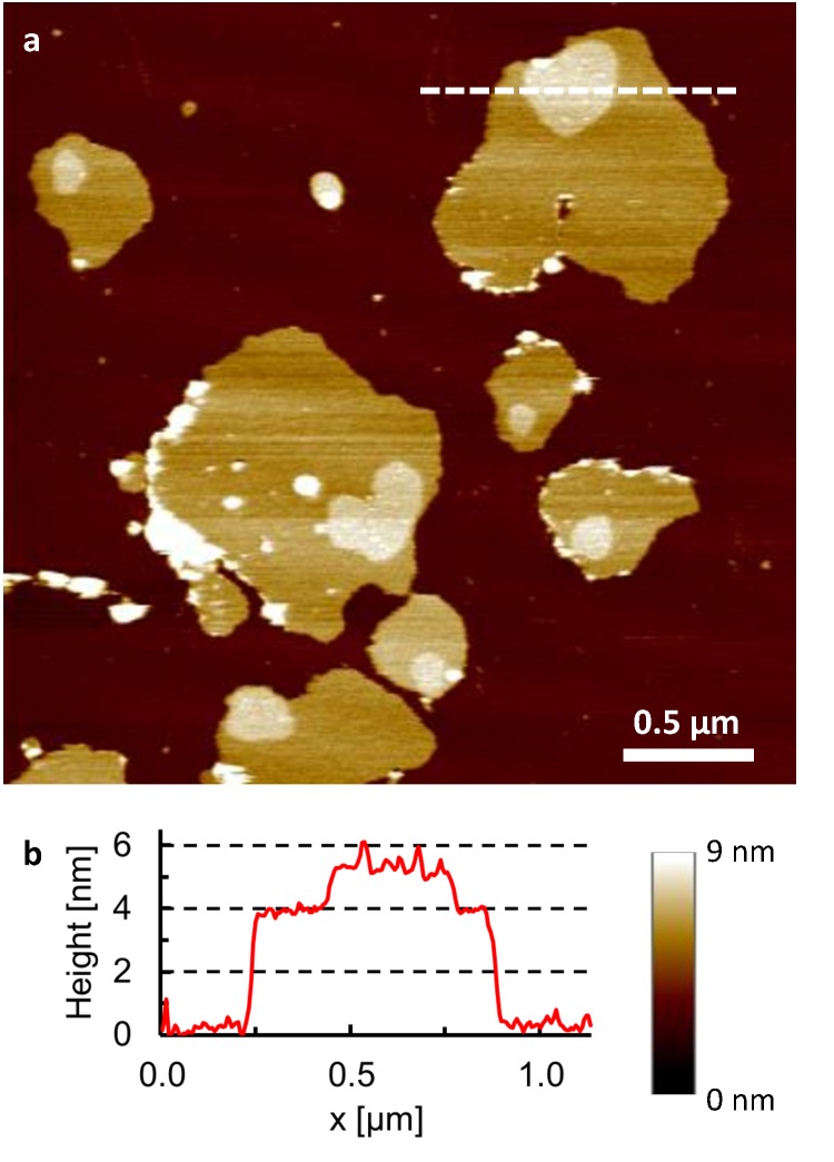
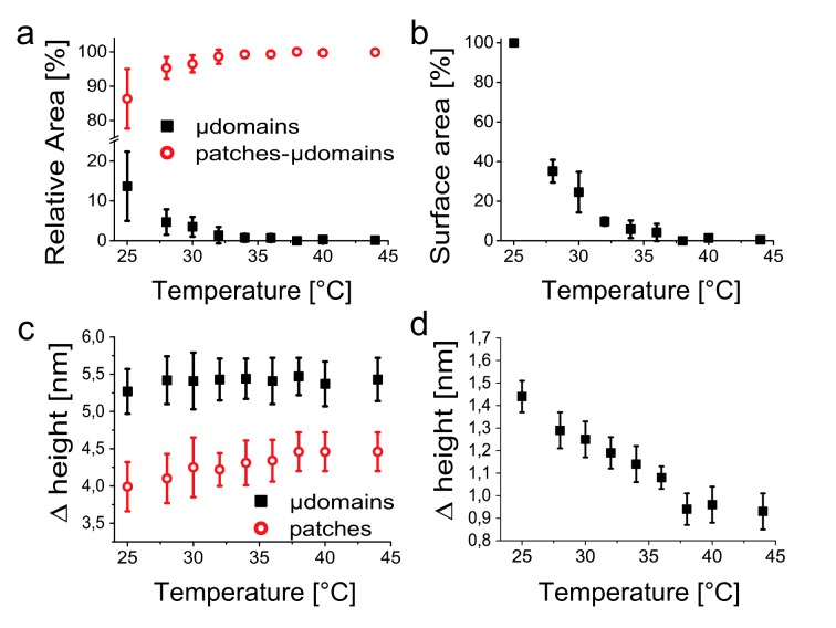
The formation of lipid microdomains ("rafts") is presumed to play an important role in various cellular functions, but their nature remains controversial. Here we report on microdomain formation in isolated, detergent-resistant membranes from MDA-MB-231 human breast cancer cells, studied by atomic force microscopy (AFM). Whereas microdomains were readily observed at room temperature, they shrunk in size and mostly disappeared at higher temperatures. This shrinking in microdomain size was accompanied by a gradual reduction of the height difference between the microdomains and the surrounding membrane, consistent with the behaviour expected for lipids that are laterally segregated in liquid ordered and liquid disordered domains. Immunolabeling experiments demonstrated that the microdomains contained flotillin-1, a protein associated with lipid rafts. The microdomains reversibly dissolved and reappeared, respectively, on heating to and cooling below temperatures around 37 °C, which is indicative of radical changes in local membrane order close to physiological temperature.


Conflict of interest statement

Competing Interests: The authors have declared that no competing interests exist.

Figures

Fig 1
Fig 1. Microdomains in detergent-resistant membranes.
a, AFM topography of detergent-resistant membranes (isolated on sucrose gradient, fraction 5) adsorbed on a mica substrate (dark brown), recorded in buffer solution at 25°C. Microdomains appear as elevated (brighter) plateaus with lateral dimensions of 100–300 nm on top of the membrane patches. b, Height profile corresponding to the white line drawn in (a).
Fig 2
Fig 2. Reversible dissolution and formation of microdomains.
a, AFM topography of isolated membrane samples in buffer solution, for a thermal cycle in which the temperature is first increased from 25°C to 30°C and then 37°C, followed by a decrease back to 25°C. Arrows indicate the sequence of the images. Vertical (color) scale for all AFM images: 9 nm, see also scale bar in Fig 1. b, Histogram of the surface area of the microdomains observed on the membrane patches in (a), demonstrating a shrinking of microdomains for higher temperatures and a full recovery at the end of the thermal cycle (25°C*). c, Histogram of the total surface area of all patches that are fully included within the frames displayed in (a), showing that the absolute changes in total patch area are small compared to the absolute changes in microdomain area over the thermal cycle. Error bars = 5% (see Methods).
Fig 3
Fig 3. Temperature dependence of microdomain size and height.
a, Plot of the relative areas of microdomains and membrane patches as a function of the temperature. b, Average of the surface area of individual microdomains as a function of temperature, normalized to the surface area of the respective microdomains at 25°C. c, Plot of the microdomain and membrane patch heights, both measured with respect to the mica, as a function of the temperature. d, Height of the microdomains measured with respect to the surrounding membrane patch surface, as a function of temperature. All data represent the mean ± SD obtained analyzing 16 different thermal cycles.
Fig 4
Fig 4. Immunolabelling of microdomains.
AFM at room temperature to detect protein content in the microdomains, before (left) and after (right) a thermal cycle (25°C -37°C- 40°C- 25°C). a, Untreated membrane patches were incubated with anti flotillin-1 antibodies. b, After 60 min of incubation, the area of the microdomains protruding from the membrane patches had increased (see differently colored circles for individual examples). c, Histogram of the surface area increase for membrane patches and microdomains after 60 min antibody incubation. d, e, f, As (a, b, c), but for membrane patches that have undergone a thermal cycle prior to the immunolabeling. Student's t-test for the difference between relative increases of patch and microdomain areas: p<0.01 in both (c) and (f). Horizontal scale bar: 1 μm; vertical (color) scale: 9 nm in the four AFM images.

References

    1. van Meer G, Voelker DR, Feigenson GW. Membrane lipids: where they are and how they behave. Nature Rev. 2008;9(2): 112–124. - PMC - PubMed
    1. Singer SJ, Nicolson GL. The fluid mosaic model of the structure of cell membranes. Science 1972;175(4023): 720–731. - PubMed
    1. Coskun U, Simons K. Membrane rafting: from apical sorting to phase segregation. FEBS Lett. 2010;584(9): 1685–1693. 10.1016/j.febslet.2009.12.043 - DOI - PubMed
    1. Sonnino S, Prinetti A. Membrane domains and the "lipid raft" concept. Curr. Med.l Chem. 2013;20(1): 4–21. - PubMed
    1. Kraft ML. Plasma membrane organization and function: moving past lipid rafts. Mol. Biol. Cell 2013;24(18): 2765–2768. 10.1091/mbc.E13-03-0165 - DOI - PMC - PubMed

Publication types