. 2015 Sep;72(9):1029-42.
doi: 10.1001/jamaneurol.2015.1285.

Longitudinal Cerebrospinal Fluid Biomarker Changes in Preclinical Alzheimer Disease During Middle Age

Affiliations

Longitudinal Cerebrospinal Fluid Biomarker Changes in Preclinical Alzheimer Disease During Middle Age

Courtney L Sutphen et al. JAMA Neurol. 2015 Sep.

Abstract

Importance: Individuals in the presymptomatic stage of Alzheimer disease (AD) are increasingly being targeted for AD secondary prevention trials. How early during the normal life span underlying AD pathologies begin to develop, their patterns of change over time, and their relationship with future cognitive decline remain to be determined.

Objective: To characterize the within-person trajectories of cerebrospinal fluid (CSF) biomarkers of AD over time and their association with changes in brain amyloid deposition and cognitive decline in cognitively normal middle-aged individuals.

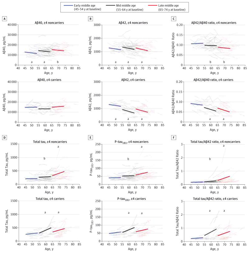
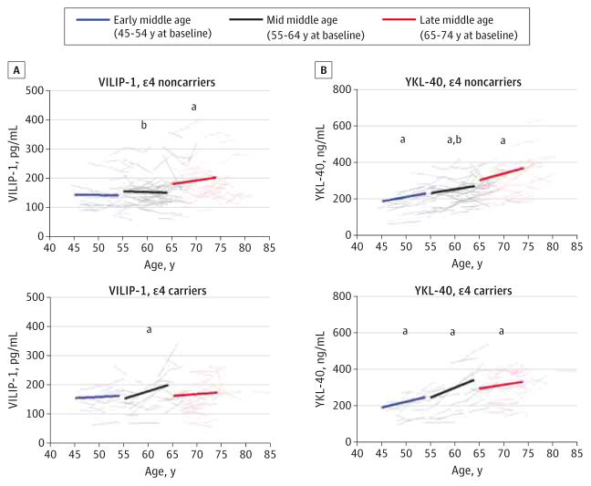
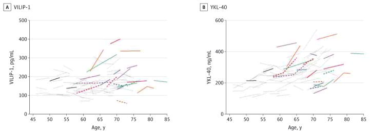
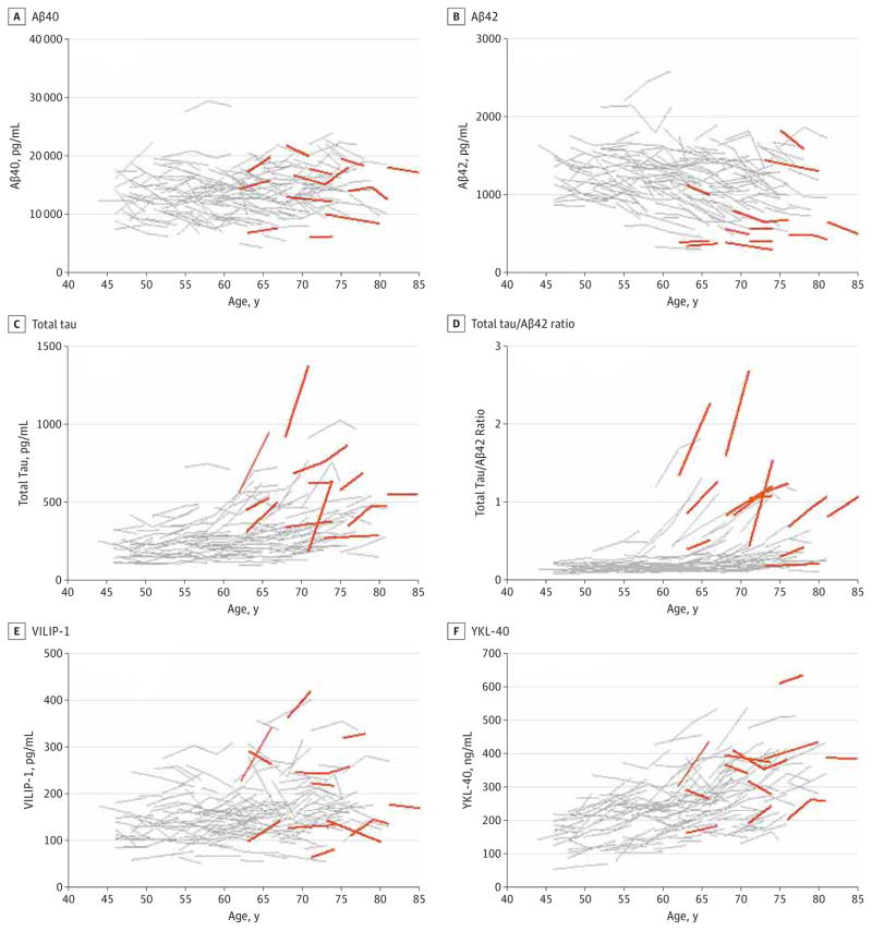
Design, setting, and participants: As part of a cohort study, cognitively normal (Clinical Dementia Rating [CDR] of 0) middle-aged research volunteers (n = 169) enrolled in the Adult Children Study at Washington University, St Louis, Missouri, had undergone serial CSF collection and longitudinal clinical assessment (mean, 6 years; range, 0.91-11.3 years) at 3-year intervals at the time of analysis, between January 2003 and November 2013. A subset (n = 74) had also undergone longitudinal amyloid positron emission tomographic imaging with Pittsburgh compound B (PiB) in the same period. Serial CSF samples were analyzed for β-amyloid 40 (Aβ40), Aβ42, total tau, tau phosphorylated at threonine 181 (P-tau181), visinin-like protein 1 (VILIP-1), and chitinase-3-like protein 1 (YKL-40). Within-person measures were plotted according to age and AD risk defined by APOE genotype (ε4 carriers vs noncarriers). Linear mixed models were used to compare estimated biomarker slopes among middle-age bins at baseline (early, 45-54 years; mid, 55-64 years; late, 65-74 years) and between risk groups. Within-person changes in CSF biomarkers were also compared with changes in cortical PiB binding and progression to a CDR higher than 0 at follow-up.

Main outcomes and measures: Changes in Aβ40, Aβ42, total tau, P-tau181, VILIP-1, and YKL-40 and, in a subset of participants, changes in cortical PiB binding.

Results: While there were no consistent longitudinal patterns in Aβ40 (P = .001-.97), longitudinal reductions in Aβ42 were observed in some individuals as early as early middle age (P ≤ .05) and low Aβ42 levels were associated with the development of cortical PiB-positive amyloid plaques (area under receiver operating characteristic curve = 0.9352; 95% CI, 0.8895-0.9808), especially in mid middle age (P < .001). Markers of neuronal injury (total tau, P-tau181, and VILIP-1) dramatically increased in some individuals in mid and late middle age (P ≤ .02), whereas the neuroinflammation marker YKL-40 increased consistently throughout middle age (P ≤ .003). These patterns were more apparent in at-risk ε4 carriers (Aβ42 in an allele dose-dependent manner) and appeared to be associated with future cognitive deficits as determined by CDR.

Conclusions and relevance: Longitudinal CSF biomarker patterns consistent with AD are first detectable during early middle age and are associated with later amyloid positivity and cognitive decline. Such measures may be useful for targeting middle-aged, asymptomatic individuals for therapeutic trials designed to prevent cognitive decline.


Conflict of interest statement

Conflict of Interest Disclosures: Dr Benzinger reported being a member of the advisory board for Eli Lilly and Co (2011); receiving research funding from Avid Radiopharmaceuticals; providing expert testimony and receiving compensation from Kujawaski and Associates (2011); and participating in clinical trials sponsored by Eli Lilly and Co, Avid Radiopharmaceuticals, and Roche. Mr Stoops reported being a shareholder of ADx NeuroSciences and BioMARIC NV. Dr Vanderstichele reported being a cofounder and shareholder of ADx NeuroSciences and founder of Biomarkable bvba. Dr Ladenson reported being named on patents related to use of VILIP-1; these are being managed by Washington University in accordance with university policy. Dr Morris reported having participated in or currently participating in clinical trials of antidementia drugs sponsored by Janssen Immunotherapy, Pfizer, Eli Lilly and Co/Avid Radiopharmaceuticals, SNIFF (Study of Nasal Insulin to Fight Forgetfulness), and A4 Study (Anti-Amyloid Treatment in Asymptomatic Alzheimer’s Disease) and serving as a consultant for Lilly USA, ISIS Pharmaceuticals, and the Charles Dana Foundation. Dr Holtzman reported being a cofounder of C2N Diagnostics LLC; serving on the scientific advisory boards of AstraZeneca, Genentech, Neurophage, and C2N Diagnostics; and serving as a consultant for Eli Lilly and Co. Washington University receives grants to the laboratory of Dr Holtzman from the Tau Consortium, Cure Alzheimer’s Fund, the JPB Foundation, Eli Lilly and Co, Janssen, and C2N Diagnostics. Dr Fagan reported serving on the scientific advisory boards of IBL International and Roche and serving as a consultant for AbbVie and Novartis. No other disclosures were reported.

Figures

Figure 1
Figure 1. Longitudinal Change in Cerebrospinal Fluid Biomarkers β-Amyloid 40 (Aβ40), Aβ42, Aβ42 to Aβ40 Ratio, Total Tau, Tau Phosphorylated at Threonine 181 (P-tau181), and Total Tau to Aβ42 Ratio During Middle Age
Estimated group slopes and within-person changes for Aβ40 (A), Aβ42 (B), Aβ42 to Aβ40 ratio (C), total tau (D), tau phosphorylated at threonine 181 (P-tau181) (E), and total tau to Aβ42 ratio (F) are shown in the 3 age bins for APOE ε4 noncarriers (top graph of each panel; n = 108 participants) and ε4 carriers (bottom graph of each panel; n = 61 participants). Annual slopes have been extrapolated to 9 years, and each slope begins at the mean baseline biomarker value from individuals in each age bin. Group baseline values and slopes represent the estimates reported in Table 1 and Table 2, respectively, for the different cohorts defined by baseline age in which biomarker concentrations were regressed on time from study entry. Data are from the INNOTEST enzyme-linked immunosorbent assay (Fujirebio Europe). aSlope significantly different from 0 (P < .05). bSlope significantly different between APOE ε4 groups within a given age group (P < .05).
Figure 2
Figure 2. Longitudinal Change in Cerebrospinal Fluid Biomarkers Visinin-Like Protein 1 (VILIP-1) and Chitinase-3-Like Protein 1 (YKL-40) During Middle Age
Estimated group slopes and within-person changes for VILIP-1 (A) and YKL-40 (B) are shown in the 3 age bins for APOE ε4 noncarriers (top graph of each panel; n = 108 participants) and ε4 carriers (bottom graph of each panel; n = 61 participants). Annual slopes have been extrapolated to 9 years, and each slope begins at the mean baseline biomarker value from individuals in each age bin. Group baseline values and slopes represent the estimates reported in Table 1 and Table 2, respectively, for the different cohorts defined by baseline age in which biomarker concentrations were regressed on time from study entry. aSlope significantly different from 0 (P < .05). bSlope significantly different between APOE ε4 groups within a given age group (P < .05).
Figure 3
Figure 3. Association Between Longitudinal Patterns of Cerebrospinal Fluid Biomarkers Cortical Pittsburgh Compound B (PiB) Standardized Uptake Value Ratio (SUVR), β-Amyloid 40 (Aβ40), Aβ42, Aβ42 to Aβ40 Ratio, and Total Tau, Cortical Amyloid, and Age
A subset (n = 74) of Adult Children Study participants had undergone longitudinal amyloid imaging via PiB positron emission tomographic imaging within 376 days (mean [SD], 84.3 [92] days) of cerebrospinal fluid collection. Biomarker measures include cortical PiB SUVR (A), Aβ40 (B), Aβ42 (C), Aβ42 to Aβ40 ratio (D), and total tau (E). The Aβ40, Aβ42, and total tau were analyzed by INNOTEST enzyme-linked immunosorbent assay (Fujirebio Europe). Being PiB positive was defined as having a mean cortical PiB SUVR higher than 1.42 and is represented by the dashed horizontal line in panel A. Gray lines indicate PiB negative at baseline and follow-up (n = 52); solid colored lines, PiB positive at both baseline and follow-up (n = 14); dashed colored lines, PiB negative at baseline but positive at follow-up (n = 6); and solid black lines, PiB negative with discordant (low) cerebrospinal fluid Aβ measures at baseline and follow-up (n = 2). Colored solid and dashed lines are each differently colored only to facilitate visual comparisons across all analytes for each PiB-positive individual.
Figure 4
Figure 4. Association Between Longitudinal Patterns of Cerebrospinal Fluid Biomarkers Visinin-Like Protein 1 (VILIP-1) and Chitinase-3-Like Protein 1 (YKL-40), Cortical Amyloid, and Age
A subset (n = 74) of Adult Children Study participants had undergone longitudinal amyloid imaging via Pittsburgh compound B (PiB) positron emission tomographic imaging within 376 days (mean [SD], 84.3 [92] days) of cerebrospinal fluid collection. Biomarker measures include VILIP-1 (A) and YKL-40 (B). Being PiB positive was defined as having a mean cortical PiB standardized uptake value ratio higher than 1.42 (see dashed horizontal line in Figure 3A). Gray lines indicate PiB negative at baseline and follow-up (n = 52); solid colored lines, PiB positive at both baseline and follow-up (n = 14); dashed colored lines, PiB negative at baseline but positive at follow-up (n = 6); and solid black lines, PiB negative with discordant (low) cerebrospinal fluid β-amyloid measures at baseline and follow-up (n = 2). Colored solid and dashed lines are each differently colored only to facilitate visual comparisons across all analytes for each PiB-positive individual.
Figure 5
Figure 5. Cerebrospinal Fluid Biomarker Trajectories in Participants Receiving a Clinical Dementia Rating Higher Than 0 at Some Point During Clinical Follow-up
Within-person trajectories of cerebrospinal fluid β-amyloid 40 (Aβ40) (A), Aβ42 (B), total tau (C), total tau to Aβ42 ratio (D), visinin-like protein 1 (VILIP-1) (E), and chitinase-3-like protein 1 (YKL-40) (F) are plotted as a function of age. The Aβ40, Aβ42, and total tau were analyzed by INNOTEST enzyme-linked immunosorbent assay (Fujirebio Europe). Fourteen individuals received a Clinical Dementia Rating of 0.5 or 1 at some point during follow-up (mean [SD], 6.55 [1.94] years; range, 4.21–10.28 years). Orange lines indicate individuals who received a Clinical Dementia Rating higher than 0 at available follow-up visits; gray lines, individuals who did not receive a Clinical Dementia Rating higher than 0.

Comment in

References

    1. James BD, Leurgans SE, Hebert LE, Scherr PA, Yaffe K, Bennett DA. Contribution of Alzheimer disease to mortality in the United States. Neurology. 2014;82(12):1045–1050. - PMC - PubMed
    1. Schneider LS, Mangialasche F, Andreasen N, et al. Clinical trials and late-stage drug development for Alzheimer’s disease: an appraisal from 1984 to 2014. J Intern Med. 2014;275(3):251–283. - PMC - PubMed
    1. Gómez-Isla T, Price JL, McKeel DW, Jr, Morris JC, Growdon JH, Hyman BT. Profound loss of layer II entorhinal cortex neurons occurs in very mild Alzheimer’s disease. J Neurosci. 1996;16(14):4491–4500. - PMC - PubMed
    1. Braak H, Braak E. Diagnostic criteria for neuropathologic assessment of Alzheimer’s disease. Neurobiol Aging. 1997;18(4 suppl):S85–S88. - PubMed
    1. Hulette CM, Welsh-Bohmer KA, Murray MG, Saunders AM, Mash DC, McIntyre LM. Neuropathological and neuropsychological changes in “normal” aging: evidence for preclinical Alzheimer disease in cognitively normal individuals. J Neuropathol Exp Neurol. 1998;57(12):1168–1174. - PubMed

Publication types

MeSH terms