. 2015 Jul 6;10(7):e0131785.
doi: 10.1371/journal.pone.0131785. eCollection 2015.

Isolation of Foreign Material-Free Endothelial Progenitor Cells Using CD31 Aptamer and Therapeutic Application for Ischemic Injury

Affiliations

Isolation of Foreign Material-Free Endothelial Progenitor Cells Using CD31 Aptamer and Therapeutic Application for Ischemic Injury

Jung Won Yoon et al. PLoS One. .

Abstract

Endothelial progenitor cells (EPCs) can be isolated from human bone marrow or peripheral blood and reportedly contribute to neovascularization. Aptamers are 40-120-mer nucleotides that bind to a specific target molecule, as antibodies do. To utilize apatmers for isolation of EPCs, in the present study, we successfully generated aptamers that recognize human CD31, an endothelial cell marker. CD31 aptamers bound to human umbilical cord blood-derived EPCs and showed specific interaction with human CD31, but not with mouse CD31. However, CD31 aptamers showed non-specific interaction with CD31-negative 293FT cells and addition of polyanionic competitor dextran sulfate eliminated non-specific interaction without affecting cell viability. From the mixture of EPCs and 293FT cells, CD31 aptamers successfully isolated EPCs with 97.6% purity and 94.2% yield, comparable to those from antibody isolation. In addition, isolated EPCs were decoupled from CD31 aptamers with a brief treatment of high concentration dextran sulfate. EPCs isolated with CD31 aptamers and subsequently decoupled from CD31 aptamers were functional and enhanced the restoration of blood flow when transplanted into a murine hindlimb ischemia model. In this study, we demonstrated isolation of foreign material-free EPCs, which can be utilized as a universal protocol in preparation of cells for therapeutic transplantation.


Conflict of interest statement

Competing Interests: The stated financial disclosure does not alter the authors' adherence to PLOS ONE policies on sharing data and materials.

Figures

Fig 1
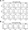
Fig 1. CD31 aptamers interact with human EPCs.
(A) Flow cytometry analysis of EPCs after individual incubation with various concentrations (0, 0.2, 2, 20, and 200 nM) of three CD31 aptamer clones (AT-1, AT-2, and AT-3, Cy5-labeled) is shown. (B) Flow cytometry analysis of EPCs after incubation with various concentrations (0, 0.2, 2, 20, and 200 nM) of control aptamers (FITC-labeled) is shown (n = 5).
Fig 2
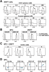
Fig 2. Dextran sulfate effectively reduces the non-specific interaction of CD31 aptamers.
(A) 293FT cells were incubated with various concentrations (0, 0.2, 2, 20, and 200 nM) of CD31 aptamer clone 1 (AT-1, Cy5-labeled) or control aptamers (FITC-labeled) and subjected to flow cytometry analysis (n = 5). (B) EPCs or 293FT cells were separately incubated with control aptamers (Ctrl AT, FITC-labeled) or CD31 aptamers (AT-1, Cy5-labeled, 200 nM) with or without 0.2 mM dextran sulfate and subjected to flow cytometry. The overlap of histograms from 0 and 0.2 mM dextran sulfate experiments is shown (n = 5). (C) The mixture of EPCs and 293FT cells was incubated with CD31 aptamers (AT-1, Cy5-labeled, 200 nM) and various concentrations (0, 0.2, 1, and 5 mM) of dextran sulfate, followed by flow cytometry analysis (n = 3). (D) EPCs were incubated with CD31 antibodies (FITC-labeled) alone, CD31 aptamers alone (AT-1, Cy5-labeled, 200 nM), or with both CD31 antibodies and CD31 aptamers and subjected to flow cytometry analysis. Two-dimensional plots are shown (n = 5).
Fig 3
Fig 3. CD31 aptamers specifically stain EPCs for visualization with fluorescence microscopy.
(A) EPCs or 293FT cells were stained with CD31 antibodies (Alexa Fluor 488-labeled) and DAPI (blue). Images were taken by confocal microscope (Olympus FluoView FV1000). (B) EPCs or 293FT cells were stained with CD31 aptamers (AT-1, biotin-labeled, 400 nM) at 37°C for 1 h with or without 0.2 mM dextran sulfate, followed by staining with streptavidin-Alexa Fluor 488 and DAPI (blue). Images were taken by confocal microscope (Olympus FluoView FV1000). Scale bar = 40 μm (n = 3).
Fig 4
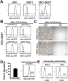
Fig 4. CD31 aptamers isolate EPCs from the mixture of EPCs and 293FT cells and EPCs are decoupled from CD31 aptamers.
(A) EPCs, 293FT cells, or the mixture of EPCs and 293FT cells were analyzed by flow cytometry after staining cells with FITC-labeled anti-human CD31 antibodies (n = 3). (B) The mixture of EPCs and 293FT cells was subjected to magnetic bead sorting using CD31 aptamers (AT-1, biotin-labeled) (upper panel) or CD31 antibodies (lower panel). (+) indicates the fraction with positive selection and (-) indicates the fraction with negative selection. Flow cytometry analysis of each fraction with CD31 antibodies (FITC-labeled) after isolation is shown (n = 3). (C) Bright field images of cultured cells in (+) fraction after magnetic bead isolation of the mixture of EPCs and 293FT cells with CD31 aptamers (upper panel) or CD31 antibodies (lower panel) are shown. Scale bar = 200 μm (n = 3). (D) Decoupling of EPCs from CD31 aptamer-EPC complexes by treatment with DxSO4. EPCs were decoupled from the CD31 aptamer-EPC complexes and recovery yield was calculated by comparing cell numbers before and after decoupling with DxSO4 (left panel) (n = 4). Flow cytometry analysis of decoupled cells with CD31 antibody is shown in the right panel. (E) Viability of EPCs before and after decoupling with DxSO4 is shown by flow cytometry analysis (n = 5).
Fig 5
Fig 5. Preparation of foreign material-free EPCs from human cord blood MNC culture by magnetic cell sorting isolation with CD31 aptamer and decoupling processes.
(A) Two-week culture of cord blood MNCs were subjected to magnetic bead isolation process with CD31 aptamers, followed by decoupling from CD31 aptamers with decoupling buffer. Bright field images (high and low magnification) of the MNCs and the EPCs decoupled from CD31 aptamers are shown. Scale bar = 100 μm. (B, C) Endothelial characteristics of the EPCs decoupled from CD31 aptamers were determined by staining with DiI-Ac-LDL (B) or lectin (Ulex europaeus agglutinin I) (C). Fluorescence images were taken by confocal microscope (Olympus FluoView FV1000). Scale bar = 40 μm. Representative data from three independent experiments are shown.
Fig 6
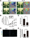
Fig 6. Foreign material-free EPCs enhance restoration of blood flow and limb salvage from ischemic injury.
(A) The foreign material-free EPCs were injected intramuscularly to injury site of nude mice with hindlimb ischemia (1×10⁶ cells/injection). Representative images of mice with laser Doppler perfusion imaging (LDPI) on day 0 and day 28 after injection with HBSS buffer (n = 6) or EPCs (n = 6) are shown. (B) Time-course quantitative analysis of blood flow by LDPI of hindlimb ischemia-induced mice injected with EPCs or HBSS is shown. (C) Statistical analysis of necrosis score on day 28 is shown. (D) Immunostaining of CD31-positive capillaries (green) in salvaged hindlimbs on day 28 after intramuscular injection of HBSS or EPCs is shown. (E) Quantitative analysis of capillary density in salvaged limbs on day 28 by counting the number of CD31-positive capillaries per high-power field (HPF) is shown. * indicates p < 0.05 vs HBSS by Student’s t-test (n = 6 for EPC and n = 6 for HBSS respectively).

Similar articles

Cited by

References

    1. Soontornworajit B, Wang Y. Nucleic acid aptamers for clinical diagnosis: cell detection and molecular imaging. Analytical and bioanalytical chemistry. 2011;399(4):1591–9. 10.1007/s00216-010-4559-x . - DOI - PubMed
    1. Ellington AD, Szostak JW. In vitro selection of RNA molecules that bind specific ligands. Nature. 1990;346(6287):818–22. 10.1038/346818a0 . - DOI - PubMed
    1. Keefe AD, Pai S, Ellington A. Aptamers as therapeutics. Nature reviews Drug discovery. 2010;9(7):537–50. 10.1038/nrd3141 . - DOI - PMC - PubMed
    1. Germer K, Leonard M, Zhang X. RNA aptamers and their therapeutic and diagnostic applications. International journal of biochemistry and molecular biology. 2013;4(1):27–40. - PMC - PubMed
    1. Sundaram P, Kurniawan H, Byrne ME, Wower J. Therapeutic RNA aptamers in clinical trials. European journal of pharmaceutical sciences: official journal of the European Federation for Pharmaceutical Sciences. 2013;48(1–2):259–71. 10.1016/j.ejps.2012.10.014 . - DOI - PubMed

Publication types

MeSH terms

Substances

LinkOut - more resources