Review
. 2015 Oct;98(2):193-207.
doi: 10.1111/mmi.13117. Epub 2015 Aug 3.

Bacterial microcompartments: widespread prokaryotic organelles for isolation and optimization of metabolic pathways

Affiliations
Review

Bacterial microcompartments: widespread prokaryotic organelles for isolation and optimization of metabolic pathways

Thomas A Bobik et al. Mol Microbiol. 2015 Oct.

Abstract

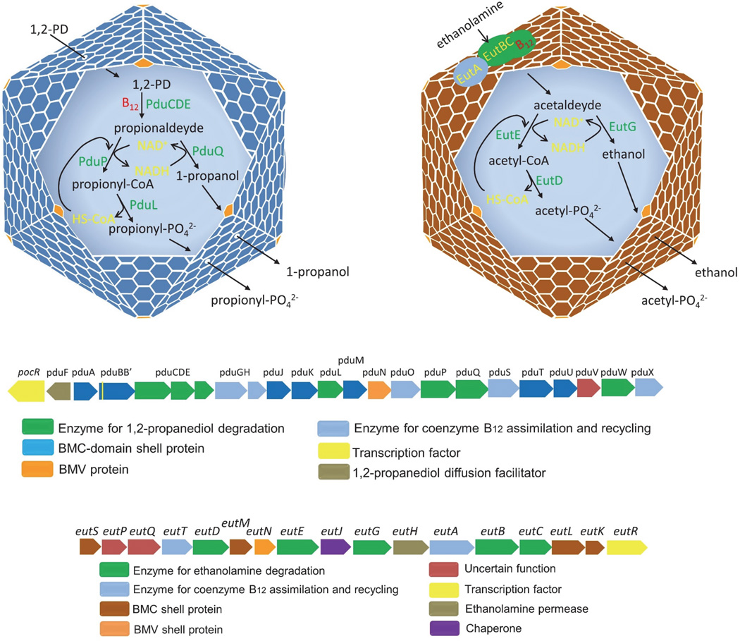
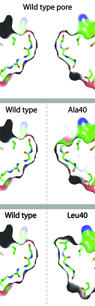
Prokaryotes use subcellular compartments for a variety of purposes. An intriguing example is a family of complex subcellular organelles known as bacterial microcompartments (MCPs). MCPs are widely distributed among bacteria and impact processes ranging from global carbon fixation to enteric pathogenesis. Overall, MCPs consist of metabolic enzymes encased within a protein shell, and their function is to optimize biochemical pathways by confining toxic or volatile metabolic intermediates. MCPs are fundamentally different from other organelles in having a complex protein shell rather than a lipid-based membrane as an outer barrier. This unusual feature raises basic questions about organelle assembly, protein targeting and metabolite transport. In this review, we discuss the three best-studied MCPs highlighting atomic-level models for shell assembly, targeting sequences that direct enzyme encapsulation, multivalent proteins that organize the lumen enzymes, the principles of metabolite movement across the shell, internal cofactor recycling, a potential system of allosteric regulation of metabolite transport and the mechanism and rationale behind the functional diversification of the proteins that form the shell. We also touch on some potential biotechnology applications of an unusual compartment designed by nature to optimize metabolic processes within a cellular context.


Figures

Figure 1
Figure 1
The carboxysome. Upper left. Model for carboxysome function. The protein shell is proposed to sequester CO2 (produced by carbonic anhydrase) in order to enhance the activity of RuBisCO as further described in the text. Upper right. Electron micrograph of purified carboxysomes from H. neapolitanus. Below: representative genetic loci encoding the two major types of carboxysomes (α and β). Below. Genes for the α-and β-carboxysome from two well-studied organisms. The details of the functions of these genes were reviewed recently (Rae et al., 2013). Lower right. Color code indicating general gene functions. (Purified carboxysomes courtesy of Gordon Cannon and Sabine Heinhorst and EM Image courtesy of Mark Yeager and Kelly Dryden).
Figure 2
Figure 2
The Pdu and Eut MCPs. Upper left: Model for the Pdu MCP. The protein shell is proposed to act as a barrier to the diffusion for propionaldehyde (produced by diol dehydratase, PduCDE) in order to minimize cellular toxicity and carbon loss as further described in the text. Upper right: An alternative model for the Eut MCP. One model for the Eut MCP is analogous to that for the Pdu MCP, but in the alternative model shown ethanolamine ammonia lyase (EutBC) is located on with the outer surface of the shell (as opposed to the lumen) where it injects acetaldehyde into the into the MCP interior. Below: genetic loci encoding the Eut and Pdu MCPs from S. enterica. The details of the functions of these genes were reviewed recently (Chowdhury et al., 2014).
Figure 3
Figure 3
Targeting of enzymes to the MCP interior by amino acid sequence extensions. Several enzymes that get compartmentalized within MCPs bear terminal sequence extensions that are responsible for binding to the interior surface of the MCP shell (Fan;Bobik, 2011, Fan et al., 2010, Fan et al., 2012, Jorda et al., 2015, Kinney et al., 2012, Choudhary et al., 2012, Lawrence et al., 2014). (A) An alignment of PduP homologues shows that sequences from bacteria that compartmentalize the enzyme within an MCP have an N-terminal extension of about 20 amino acids (adapted from (Fan et al., 2010)). (B) Analogous extensions are present on enzymes from other MCP systems (Fan;Bobik, 2011, Fan et al., 2010, Fan et al., 2012, Jorda et al., 2015, Kinney et al., 2012, Choudhary et al., 2012, Lawrence et al., 2014), adapted from (Yeates et al., 2013). (C) In the absence of crystal structures of bound complexes, computational docking models have proposed a mode of specific docking between the targeting tails and the interior surface of BMC shell proteins (adapted from (Yeates et al., 2013).
Figure 4
Figure 4
The structure and assembly of shell proteins in bacterial MCPs. Based on crystallographic and electron microscopy studies, an atomic-level model for the shells of bacterial MCPs has been developed (Tanaka et al., 2008, Yeates et al., 2011). The MCP shell proteins exist in several variations that perform varied roles in the MCP shell. Structures are shown for several examples across diverse MCP types. Shell proteins belonging to the BMC family are shown in different bluish shades from violet to blue-green. They form hexagonal tiles. A bacterial microcompartment vertex (BMV) protein, which is distinct from the BMC family is shown in red. Proteins from this family have pentagonal structures and are presumed to occupy vertices in the intact MCP. An idealized geometric model of an assembled MCP is shown. Except for some alpha carboxysomes, most MCPs are more irregular in shape.
Figure 5
Figure 5
Small mutational changes in the pore of PduA that affect transport selectivity. The images show a thin slice through the central pore of the PduA hexamer. Atoms are colored by type (carbon:green, oxygen:red, nitrogen:blue). (top) Wild-type PduA, with the side chain of serine 40 occupying the top region of the cutaway. (middle and bottom) Juxtaposed views of the wild type pore on the left with mutant pores on the right. The removal of a hydroxyl group by replacing Ser40 with an Ala40 mutation allows escape of the toxic propionaldehyde, but normal 1,2-propanediol entry. The hydrophobic side chain in the Leu40 mutant impairs uptake of the 1,2-propanediol substrate.

References

    1. Abdul-Rahman F, Petit E, Blanchard JL. The distribution of polyhedral bacterial microcompartments suggests frequent horizontal transfer and operon reassembly. J. Phylogen. Evolution. Biol. 2013;1
    1. Axen SD, Erbilgin O, Kerfeld CA. A taxonomy of bacterial microcompartment loci constructed by a novel scoring method. PLoS Comput. Biol. 2014;10:e1003898. - PMC - PubMed
    1. Badger MR, Hanson D, Price GD. Evolution and diversity of CO2 concentrating mechanisms in cyanobacteria. Functional Plant Biology. 2002;29:161–173. - PubMed
    1. Baker SH, Williams DS, Aldrich HC, Gambrell AC, Shively JM. Identification and localization of the carboxysome peptide Csos3 and its corresponding gene in Thiobacillus neapolitanus. Arch Microbiol. 2000;173:278–283. - PubMed
    1. Bobik TA, Xu Y, Jeter RM, Otto KE, Roth JR. Propanediol utilization genes (pdu) of Salmonella typhimurium: three genes for the propanediol dehydratase. J Bacteriol. 1997;179:6633–6639. - PMC - PubMed

Publication types

Substances