

Primary microRNA processing is functionally coupled to RNAP II transcription in vitro
- PMID: 26149087
- PMCID: PMC4493704
- DOI: 10.1038/srep11992
Primary microRNA processing is functionally coupled to RNAP II transcription in vitro
Abstract
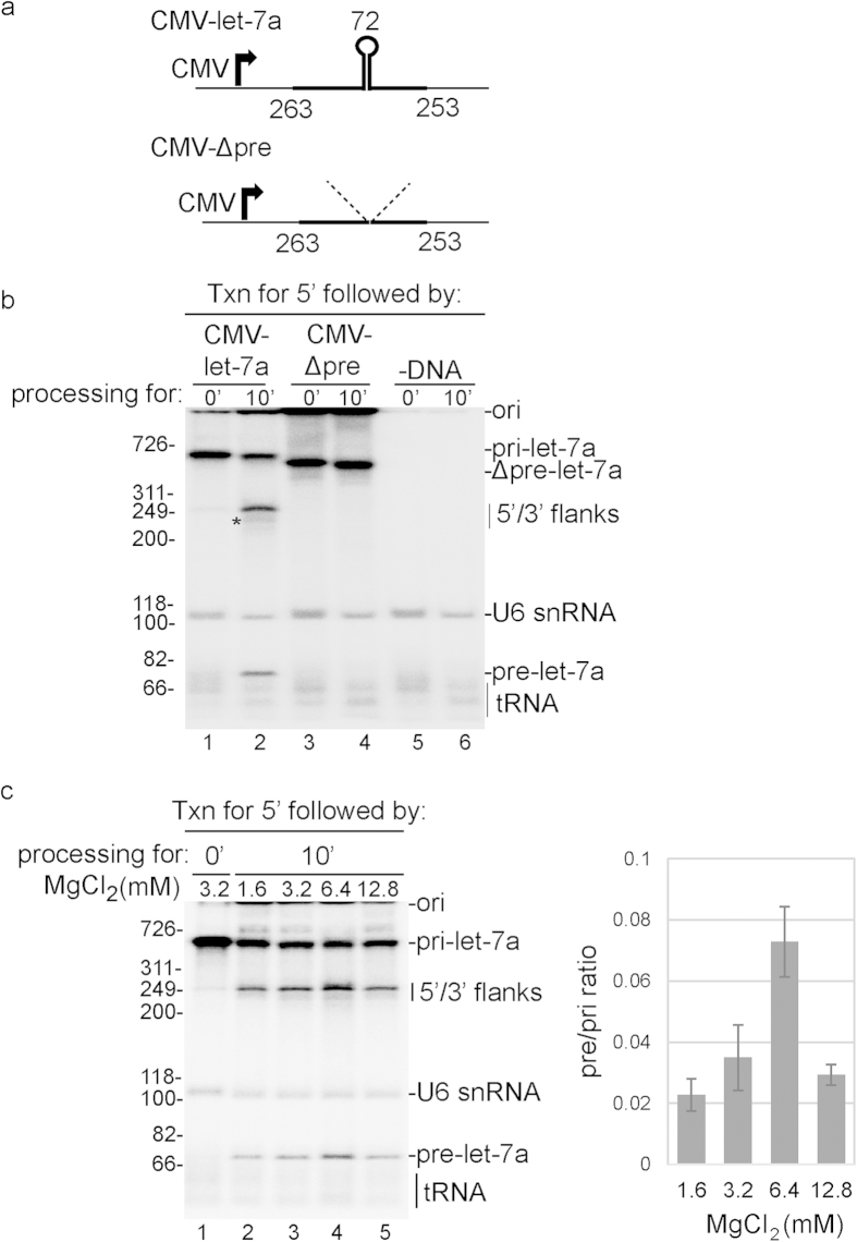
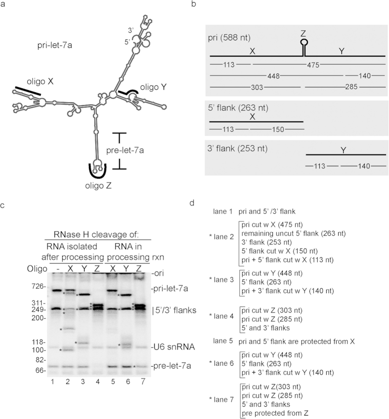
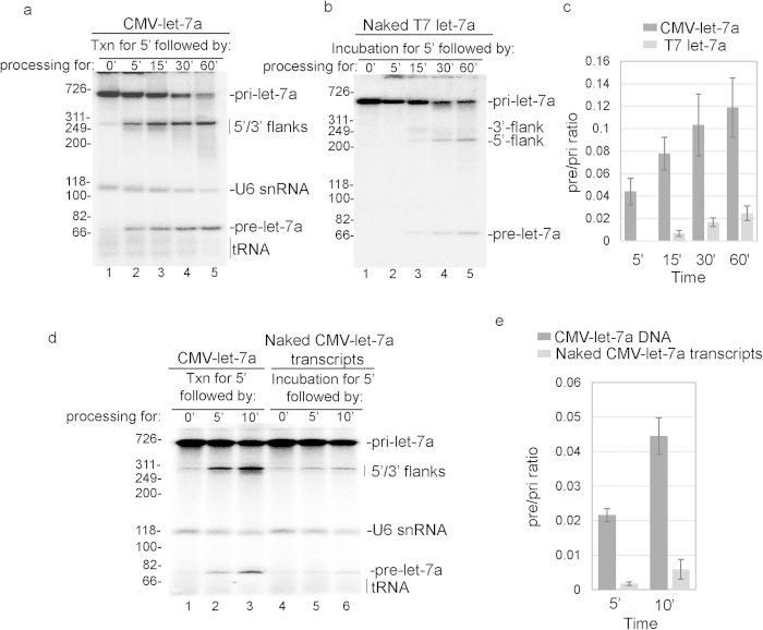
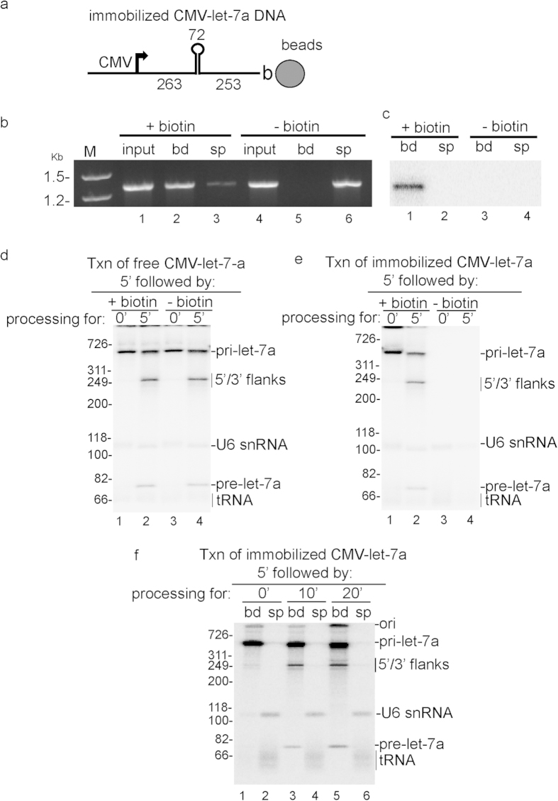
Previous studies in vivo reported that processing of primary microRNA (pri-miRNA) is coupled to transcription by RNA polymerase II (RNAP II) and can occur co-transcriptionally. Here we have established a robust in vivo system in which pri-miRNA is transcribed by RNAP II and processed to pre-miRNA in HeLa cell nuclear extracts. We show that both the kinetics and efficiency of pri-miRNA processing are dramatically enhanced in this system compared to that of the corresponding naked pri-miRNA. Moreover, this enhancement is general as it occurs with multiple pri-miRNAs. We also show that nascent pri-miRNA is efficiently processed before it is released from the DNA template. Together, our work directly demonstrates that transcription and pri-miRNA processing are functionally coupled and establishes the first in vivo model systems for this functional coupling and for co-transcriptional processing.
Figures
Similar articles
-
In Vitro System for Coupling RNAP II Transcription to Primary microRNA Processing and a Three-Way System for RNAP II Transcription/Splicing/microRNA Processing.Methods Mol Biol. 2018;1823:43-50. doi: 10.1007/978-1-4939-8624-8_4. Methods Mol Biol. 2018. PMID: 29959672 Free PMC article.
-
New insights into pri-miRNA processing and accumulation in plants.Wiley Interdiscip Rev RNA. 2015 Sep-Oct;6(5):533-45. doi: 10.1002/wrna.1292. Epub 2015 Jun 29. Wiley Interdiscip Rev RNA. 2015. PMID: 26119101 Review.
-
Human microRNAs are processed from capped, polyadenylated transcripts that can also function as mRNAs.RNA. 2004 Dec;10(12):1957-66. doi: 10.1261/rna.7135204. Epub 2004 Nov 3. RNA. 2004. PMID: 15525708 Free PMC article.
-
Subnuclear compartmentalization of transiently expressed polyadenylated pri-microRNAs: processing at transcription sites or accumulation in SC35 foci.Cell Cycle. 2009 Feb 1;8(3):345-56. doi: 10.4161/cc.8.3.7494. Epub 2009 Feb 21. Cell Cycle. 2009. PMID: 19177009 Free PMC article.
-
Isolation and identification of gene-specific microRNAs.Methods Mol Biol. 2006;342:313-20. doi: 10.1385/1-59745-123-1:313. Methods Mol Biol. 2006. PMID: 16957385 Review.
Cited by
-
Microprocessor Recruitment to Elongating RNA Polymerase II Is Required for Differential Expression of MicroRNAs.Cell Rep. 2017 Sep 26;20(13):3123-3134. doi: 10.1016/j.celrep.2017.09.010. Cell Rep. 2017. PMID: 28954229 Free PMC article.
-
Analysis of primary microRNA loci from nascent transcriptomes reveals regulatory domains governed by chromatin architecture.Nucleic Acids Res. 2017 Sep 29;45(17):9837-9849. doi: 10.1093/nar/gkx680. Nucleic Acids Res. 2017. PMID: 28973462 Free PMC article.
-
The Potential Role of miRNAs as Predictive Biomarkers in Neurodevelopmental Disorders.J Mol Neurosci. 2021 Jul;71(7):1338-1355. doi: 10.1007/s12031-021-01825-7. Epub 2021 Mar 27. J Mol Neurosci. 2021. PMID: 33774758 Review.
-
The Implications of Noncoding RNAs in the Evolution and Progression of Nonalcoholic Fatty Liver Disease (NAFLD)-Related HCC.Int J Mol Sci. 2022 Oct 15;23(20):12370. doi: 10.3390/ijms232012370. Int J Mol Sci. 2022. PMID: 36293225 Free PMC article. Review.
-
Evidence that C9ORF72 Dipeptide Repeat Proteins Associate with U2 snRNP to Cause Mis-splicing in ALS/FTD Patients.Cell Rep. 2017 Jun 13;19(11):2244-2256. doi: 10.1016/j.celrep.2017.05.056. Cell Rep. 2017. PMID: 28614712 Free PMC article.
References
-
- Greenleaf A. L. Positive patches and negative noodles: linking RNA processing to transcription? Trends Biochem Sci 18, 117–9 (1993). - PubMed
-
- Maniatis T. & Reed R. An extensive network of coupling among gene expression machines. Nature 416, 499–506. (2002). - PubMed
-
- Prasanth K. V. & Spector D. L. Eukaryotic regulatory RNAs: an answer to the ‘genome complexity’ conundrum. Genes Dev 21, 11–42 (2007). - PubMed
Publication types
MeSH terms
Substances
Grants and funding
LinkOut - more resources
Full Text Sources
Other Literature Sources
Molecular Biology Databases
Research Materials
