. 2015 Sep;35(18):3145-62.
doi: 10.1128/MCB.00332-15. Epub 2015 Jul 6.

Ezrin Binds to DEAD-Box RNA Helicase DDX3 and Regulates Its Function and Protein Level

Affiliations

Ezrin Binds to DEAD-Box RNA Helicase DDX3 and Regulates Its Function and Protein Level

Haydar Çelik et al. Mol Cell Biol. 2015 Sep.

Abstract

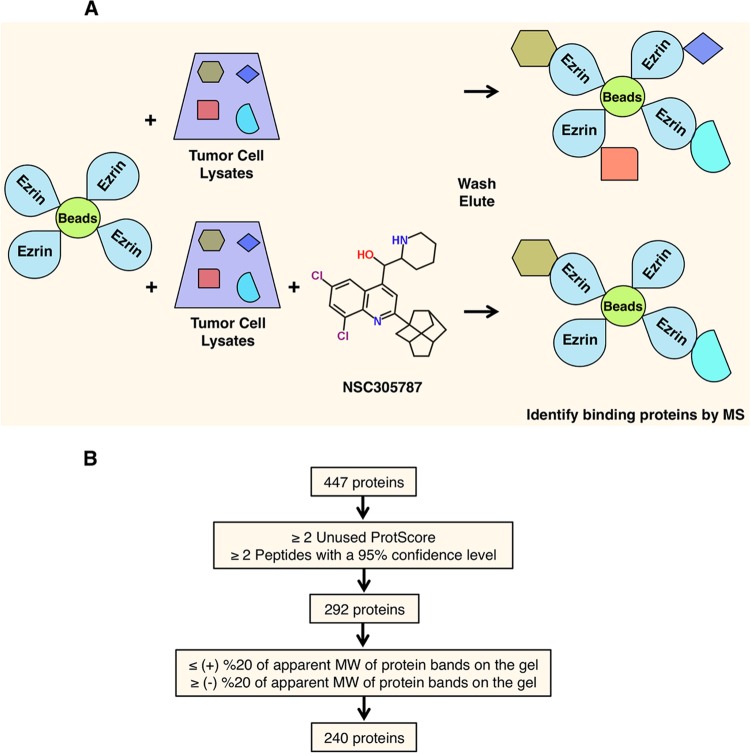
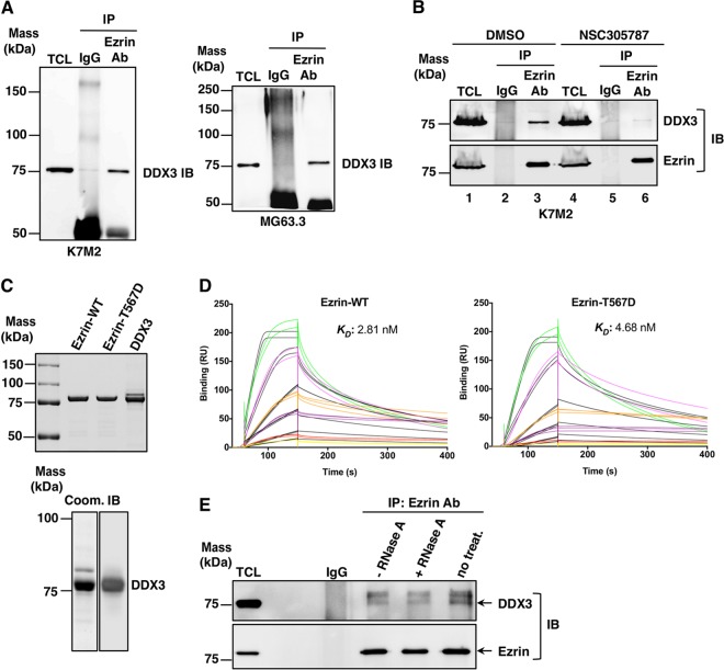
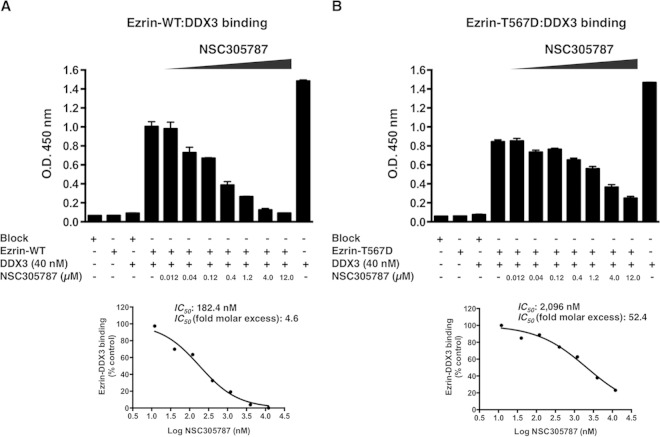
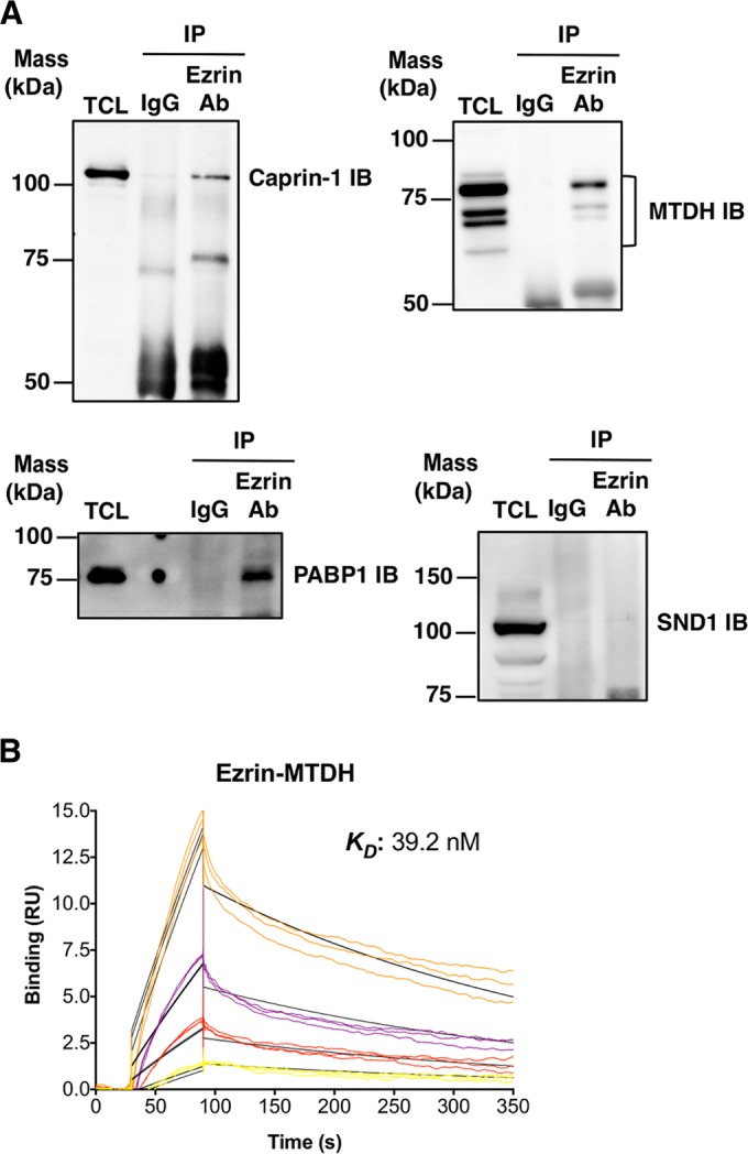
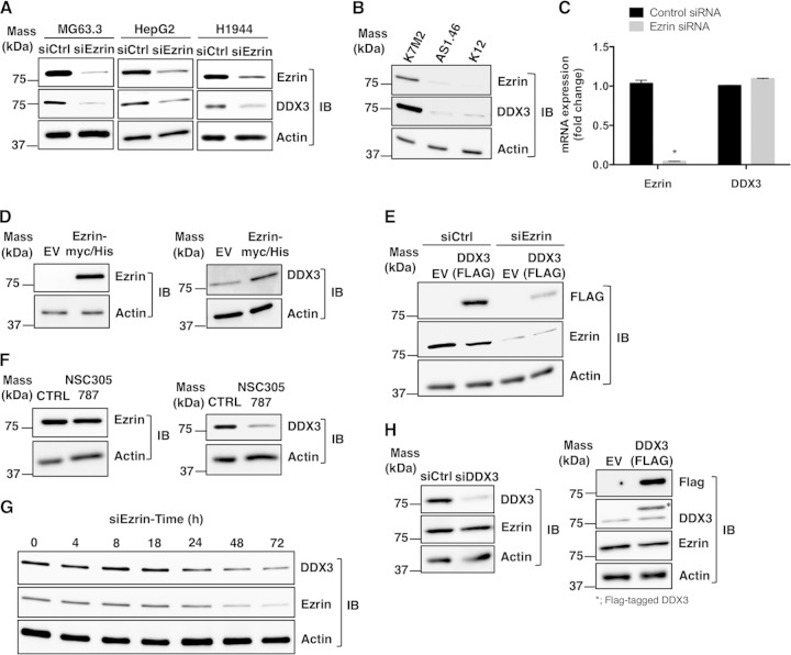
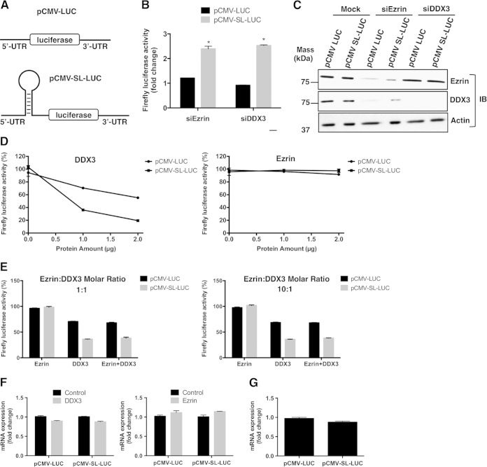
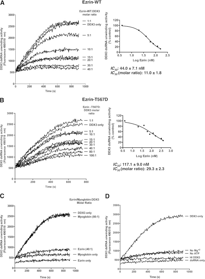
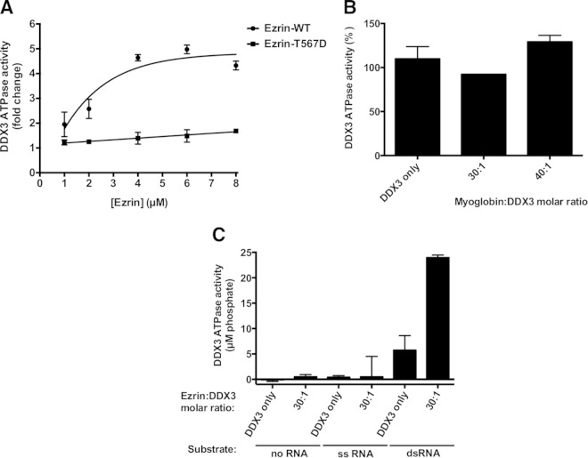
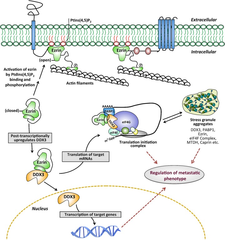
Ezrin is a key regulator of cancer metastasis that links the extracellular matrix to the actin cytoskeleton and regulates cell morphology and motility. We discovered a small-molecule inhibitor, NSC305787, that directly binds to ezrin and inhibits its function. In this study, we used a nano-liquid chromatography-tandem mass spectrometry (nano-LC-MS-MS)-based proteomic approach to identify ezrin-interacting proteins that are competed away by NSC305787. A large number of the proteins that interact with ezrin were implicated in protein translation and stress granule dynamics. We validated direct interaction between ezrin and the RNA helicase DDX3, and NSC305787 blocked this interaction. Downregulation or long-term pharmacological inhibition of ezrin led to reduced DDX3 protein levels without changes in DDX3 mRNA. Ectopic overexpression of ezrin in low-ezrin-expressing osteosarcoma cells caused a notable increase in DDX3 protein levels. Ezrin inhibited the RNA helicase activity of DDX3 but increased its ATPase activity. Our data suggest that ezrin controls the translation of mRNAs preferentially with a structured 5' untranslated region, at least in part, by sustaining the protein level of DDX3 and/or regulating its function. Therefore, our findings suggest a novel function for ezrin in regulation of gene translation that is distinct from its canonical role as a cytoskeletal scaffold at the cell membrane.


Figures

FIG 1
FIG 1
Identification of candidate NSC305787-competed ezrin-binding proteins. (A) Schematic diagram describing an affinity pulldown coupled with an MS-MS approach for identification of ezrin-interacting proteins that can be competed away by NSC305787. Recombinant ezrin was purified and coupled with CNBr-activated Sepharose 4B beads. The ezrin-coated beads were then used as bait on total cell lysates from K7M2 mouse OS cells in the presence of either NSC305787 or vehicle control. The proteins bound to the beads were eluted by boiling samples in SDS-PAGE sample buffer. Proteins were then run on an SDS-PAGE gel and stained with Coomassie brilliant blue. Protein bands that were competed away with NSC305787 were analyzed by nano-LC–MS-MS. (B) Schematic flow diagram for analysis of the raw data obtained from MS. Through stepwise filtering, a total of 240 potential ezrin-interacting proteins that can be competed away by NSC305787 were identified. Proteins identified by MS-MS were first filtered based on the elimination of proteins that were not identified by at least two unique peptides with a confidence level of ≥95% and with a ProteinPilot Unused ProtScore of ≥2 (99% confidence level). Further subtraction of proteins was then based on the removal of proteins whose predicted molecular weights differed by more than 20% from the molecular weights of the corresponding bands on the gel.
FIG 2
FIG 2
Ezrin interacts with DDX3, and the antiezrin compound NSC305787 inhibits this interaction. (A) Interaction of ezrin with DDX3 in K7M2 mouse (left) and MG63.3 human (right) OS total cell lysates (TCL) by co-IP experiments. Protein complexes were immunoprecipitated from the cell lysates with antiezrin antibody (Ab) or negative-control total mouse IgG and immunoblotted (IB) with anti-DDX3 antibody. (B) K7M2 cells were treated with 3.0 μM NSC305787 for 8 h. Total cell lysates were immunoprecipitated with control IgG or an ezrin antibody, which was followed by Western blotting for ezrin and DDX3. (C) (Top) Recombinant wild-type ezrin and the phosphomimicking ezrin T567D mutant were expressed in bacteria and purified by cation-exchange chromatography on an SP Sepharose column, followed by adsorption chromatography on a heparin column. DDX3 protein was expressed in baculovirus-infected insect cells and purified by Ni2+ affinity column chromatography. Approximately 1.5 μg of each protein was run on a gel and stained with Coomassie blue. (Bottom) A purified DDX3 sample (1.5 μg) was run on a gel and stained with Coomassie blue (Coom.) (left), or 250 ng of purified DDX3 was run on a gel and transferred to a membrane for Western blot analysis using anti-DDX3 antibody (right). (D) Recombinant wild-type ezrin or the phosphomimicking ezrin mutant was immobilized on a CM5 chip in Biacore T-200. DDX3 was injected over the chip surface at six different concentrations (1.25, 2.5, 5.0, 10, 20, and 40 nM) in triplicate. The colored lines show real data, and the black lines represent curves fitted for a 1:1 binding model. (E) Equal amounts of lysates from K7M2 mouse OS cells were subjected to co-IP with antiezrin antibody. The resulting coprecipitates were treated with 0.5 mg/ml RNase A at 37°C for 30 min. −RNase A and no treat., samples that were incubated in the absence of RNase A for 30 min at 37°C or at 4°C, respectively. Immunoblotting was performed with anti-DDX3 and antiezrin antibodies.
FIG 3
FIG 3
ELISA demonstrating inhibition of the ezrin-DDX3 protein interaction by NSC305787. NSC305787 was much more effective in competing away the binding of DDX3 to wild-type ezrin than that to the ezrin T567D phosphomimicking mutant. (A) The surface of an ELISA plate was coated with wild-type ezrin protein (300 ng/well). Binding of DDX3 to the immobilized ezrin protein was detected using anti-DDX3 antiserum, followed by a secondary antibody coupled to horseradish peroxidase and a chromogenic substrate. ELISA wells coated with BSA served as negative controls (far-left bar). Additional negative controls included wells coated with ezrin protein alone (second bar) and wells coated with BSA alone followed by incubation with DDX3 protein (third bar). For the positive control, ELISA wells were coated with DDX3 protein alone (far-right bar). The inhibitory activity of NSC305787 was tested using a range of concentrations from 0.012 μM to 12.0 μM. Inhibition of the ezrin-DDX3 interaction led to a reduction in the color signal. The graph below shows the IC50 curve. (B) ELISA performed as for panel A, except that the purified ezrin T567D phosphomimicking mutant was used in place of the wild-type protein.
FIG 4
FIG 4
Ezrin interacts with caprin-1, MTDH, and PABP1. (A) The ability of ezrin to interact with caprin-1, MTDH, PABP1, and SND1 was tested by co-IP experiments in mouse K7M2 OS total cell lysates. Protein complexes were immunoprecipitated from the cell lysates with antiezrin antibody or negative-control total mouse IgG, followed by immunoblotting with anti-caprin-1, anti-MTHD, anti-PABP1, and anti-SND1 antibodies. (B) For SPR studies, recombinant human ezrin was immobilized on a CM5 chip in Biacore T-200. Recombinant human MTDH was injected over the chip surface at four different concentrations (0.63, 1.25, 2.50, and 5.0 nM) in triplicate. The colored lines show real data, and the black lines represent curves fitted for a 1:1 binding model.
FIG 5
FIG 5
Ezrin regulates the DDX3 protein level. (A) Expression of ezrin protein was inhibited by siRNA in human OS (MG63.3), hepatocellular cancer (HepG2), and lung cancer (H1944) cell lines, which resulted in reduced DDX3 expression. Protein levels were detected by Western blotting of total cell lysates. (B) K12 is a mouse OS cell line, and K7M2 is a subclone of K12 with high ezrin expression and high metastatic potential. Ezrin expression in K7M2 was inhibited by stable antisense oligonucleotides (labeled as AS1.46 cells). The expression levels of ezrin, DDX3, and actin proteins were determined by Western blotting. (C) Ezrin and DDX3 mRNA levels were measured by real-time PCR in MG63.3 cells from panel A, and the results were normalized to 18S rRNA levels and expressed as fold change over the control group (*, P < 0.0001; Student's t test). The values are presented as the means and standard deviations of the results of triplicate experiments. (D) Low-ezrin-expressing K12 mouse OS cells were transfected with either an empty vector (EV) or a cDNA coding for wild-type ezrin containing a Myc-His tag. The protein levels of ezrin, DDX3, and actin were detected by Western blotting after 48 h. (E) MG63.3 cells were transfected with a cDNA coding for DDX3 containing a FLAG tag or a negative-control EV. Ezrin expression was inhibited by siRNA, and the level of expression of DDX3 from the vector was determined by Western blotting using anti-FLAG antibody. (F) K7M2 cells were treated with 2.0 μM NSC305787 or vehicle control for 4 days, and the expression levels of ezrin, DDX3, and actin were determined by Western blotting. (G) The expression levels of DDX3 in MG63.3 cells were determined at 0 h, 4 h, 8 h, 18 h, 24 h, 48 h, and 72 h after transfection of cells with siRNA for ezrin. (H) MG63.3 cells were transfected with either an siRNA targeting endogenous DDX3 (left) or a cDNA coding for DDX3 containing a FLAG tag (right). The expression level of ezrin was determined by Western blotting using antiezrin antibody. The asterisk indicates the Flag-tagged exogenous DDX3.
FIG 6
FIG 6
Ezrin modulates translation of structured mRNAs through DDX3 in transfected cells, and DDX3 preferentially inhibits translation of 5′ UTR structured mRNA in vitro. (A) MG63.3 human OS cells were transfected with constructs designed to represent a nonstructured (pCMV-LUC) or a weakly translated gene with a structured (pCMV-SL-LUC) 5′ untranslated region adjacent to the reporter fLUC gene. Cells were also transfected with anti-DDX3 or antiezrin siRNAs. (B) Fold changes in normalized firefly luciferase activities from both structured and nonstructured luciferase mRNAs in ezrin- and DDX3-depleted cells with respect to mock-transfected cells. The firefly luciferase data were normalized to total protein (*, P < 0.0001; Student's t test). The values are presented as the means and standard deviations of the results of triplicate experiments. The graph shows a representative of three independent experiments. (C) Western blots showing ezrin and DDX3 protein levels from panel B. (D) Rabbit reticulocyte lysates containing pCMV-LUC or pCMV-SL-LUC reporter constructs carrying luciferase mRNAs with a nonstructured or stem-loop-structured 5′ UTR, respectively, were incubated with increasing amounts of DDX3 (1 and 2 μg, corresponding to 0.27 and 0.53 μM, respectively) or ezrin (1 and 2 μg, corresponding to 0.25 and 0.50 μM, respectively) under conditions for coupled transcription-translation. The translation was performed in a 50-μl reaction volume at 30°C for 90 min, as described in Materials and Methods. The amount of functionally active luciferase protein synthesized from a non-stem-loop- or stem-loop-structured mRNA was measured in a luminometer. The values are presented as the means ± standard deviations of triplicate determinations. (E) Reactions were set up as for panel D, except the effect of ezrin on luciferase expression was tested in the presence of DDX3. The graph on the left shows the translation reactions for pCMV-LUC and pCMV-SL-LUC performed in the presence of 1.0 μg ezrin alone (two left bars), 1.0 μg DDX3 alone (two middle bars), and both 1.0 μg ezrin and 1.0 μg DDX3 (two right bars). The graph on the right shows translation reactions for pCMV-LUC and pCMV-SL-LUC performed in the presence of 10.0 μg ezrin alone (two left bars), 1.0 μg DDX3 alone (two middle bars), and both 10.0 μg ezrin and 1.0 μg DDX3 (two right bars). (F) In vitro-coupled transcription-translation reactions were set up as for panel D with either recombinant DDX3 (1 μg) or ezrin (1 μg). After a 90-min incubation period at 30°C, total RNA was isolated as described in Materials and Methods. Luciferase mRNA levels were measured by real-time PCR, and the results were normalized to the 18S rRNA levels. The values are presented as the means and standard deviations of triplicate determinations. (G) Total RNA was isolated as described for panel E from in vitro-coupled transcription-translation reactions with rabbit reticulocyte lysates containing pCMV-LUC or pCMV-SL-LUC reporter constructs in the absence of any recombinant protein. Luciferase mRNA levels were measured by real-time PCR, and the results were normalized to 18S rRNA levels. The values are presented as the means and standard deviations of triplicate determinations.
FIG 7
FIG 7
Wild-type ezrin inhibits RNA helicase activity of DDX3 more effectively than the phosphomimicking ezrin T567D mutant in a concentration-dependent manner. (A) The effect of ezrin on DDX3 helicase activity was tested by a FRET-based reporter assay in real time. Double-stranded RNA with a 3′ overhang consisting of a 5′ fluorophore-labeled reporter strand and a 3′ quencher-labeled strand was used as the substrate. Reaction mixtures containing 4 nM recombinant DDX3 and 20 nM dsRNA with a 3′ overhang were incubated with increasing amounts of recombinant wild-type ezrin (4 to 160 nM, corresponding to 1- to 40-fold molar excesses of ezrin over DDX3). The measured fluorescence intensity indicates the amount of single-stranded RNA product formation. (Right) Nonlinear curve fitting of the concentration-response data to calculate the IC50 of wild-type ezrin on helicase activity. (B) The RNA duplex-unwinding activity of DDX3 was measured under the same conditions as described above, except instead of wild-type ezrin, the phosphomimicking ezrin T567D mutant (4 to 400 nM, corresponding to 1- to 100-fold molar excesses of ezrin over DDX3) was used in the reaction mixtures. Both graphs are representative of the results of three independent experiments. The calculated IC50 data for both wild-type ezrin and the phosphomimicking ezrin mutant represent the means ± standard deviations from three independent experiments. (C) Ezrin at 40-fold molar excess concentration over DDX3 caused almost complete inhibition of DDX3 helicase activity, whereas myoglobin as a negative-control protein at 80-fold molar excess did not inhibit the helicase activity. Ezrin or myoglobin itself did not produce any change in fluorescence readings over the time course of the experiment. (D) When ATP and Mg2+ ions were removed from the reaction mixture, no helicase activity was observed. Heat-inactivated DDX3 was also unable to catalyze the unwinding of dsRNA. The graphs show representatives of three independent experiments.
FIG 8
FIG 8
Wild-type ezrin stimulates ATPase activity of DDX3 more effectively than the phosphomimicking ezrin T567D mutant in a concentration-dependent manner. (A) The effect of ezrin on DDX3 ATPase activity was determined using a fixed-type assay by measuring the amount of free phosphate generated during the hydrolysis of ATP, as described in Materials and Methods. Reaction mixtures containing 200 nM DDX3 and 400 nM dsRNA with a 3′ overhang were incubated with increasing amounts of either recombinant wild-type ezrin or the phosphomimicking ezrin T567D mutant (1 to 8 μM, corresponding to 5- to 40-fold molar excesses of ezrin over DDX3). The graphs are representative of the results of three independent experiments. (B) When myoglobin was added to the reaction mixtures as a negative-control protein at increasing concentrations corresponding to 30- and 40-fold molar excesses over the amount of DDX3, no apparent change in enzyme activity was observed, whereas ezrin at the same concentrations significantly stimulated DDX3 ATPase activity. The results are expressed as means and standard deviations of duplicate determinations. (C) Reaction mixtures containing 200 nM DDX3 were incubated with 6 μM recombinant wild-type ezrin (corresponding to a 30-fold molar excess of ezrin over DDX3) without any RNA substrate or in the presence of 400 nM either dsRNA with a 3′ overhang or its complementary, longer ssRNA. The sequence of the dsRNA is given in Materials and Methods.
FIG 9
FIG 9
Working model for ezrin and DDX3 functions. The model demonstrates a novel role for ezrin in regulating transcription and translation independently of its membrane-localized open conformation. We propose that ezrin and DDX3 are the components of an mRNP complex associated with translation initiation machinery and stress granule aggregates. In this model, ezrin functions as a regulator of mRNP homeostasis required for transitions between translationally active (translation initiation complex) and inactive (stress granule) states under normal or oncogenic/stress conditions.

Similar articles

Cited by

References

    1. Takeuchi K, Kawashima A, Nagafuchi A, Tsukita S. 1994. Structural diversity of band 4.1 superfamily members. J Cell Sci 107:1921–1928. - PubMed
    1. Tsukita S, Yonemura S. 1997. ERM (ezrin/radixin/moesin) family: from cytoskeleton to signal transduction. Curr Opin Cell Biol 9:70–75. doi:10.1016/S0955-0674(97)80154-8. - DOI - PubMed
    1. Bretscher A, Edwards K, Fehon RG. 2002. ERM proteins and merlin: integrators at the cell cortex. Nat Rev Mol Cell Biol 3:586–599. doi:10.1038/nrm882. - DOI - PubMed
    1. Fehon RG, McClatchey AI, Bretscher A. 2010. Organizing the cell cortex: the role of ERM proteins. Nat Rev Mol Cell Biol 11:276–287. doi:10.1038/nrm2866. - DOI - PMC - PubMed
    1. Matsui T, Maeda M, Doi Y, Yonemura S, Amano M, Kaibuchi K, Tsukita S, Tsukita S. 1998. Rho-kinase phosphorylates COOH-terminal threonines of ezrin/radixin/moesin (ERM) proteins and regulates their head-to-tail association. J Cell Biol 140:647–657. doi:10.1083/jcb.140.3.647. - DOI - PMC - PubMed

Publication types

MeSH terms

LinkOut - more resources