. 2015 Sep;35(18):3200-11.
doi: 10.1128/MCB.00107-15. Epub 2015 Jul 6.

Genome-Wide Screens in Saccharomyces cerevisiae Highlight a Role for Cardiolipin in Biogenesis of Mitochondrial Outer Membrane Multispan Proteins

Affiliations

Genome-Wide Screens in Saccharomyces cerevisiae Highlight a Role for Cardiolipin in Biogenesis of Mitochondrial Outer Membrane Multispan Proteins

Julia Sauerwald et al. Mol Cell Biol. 2015 Sep.

Abstract

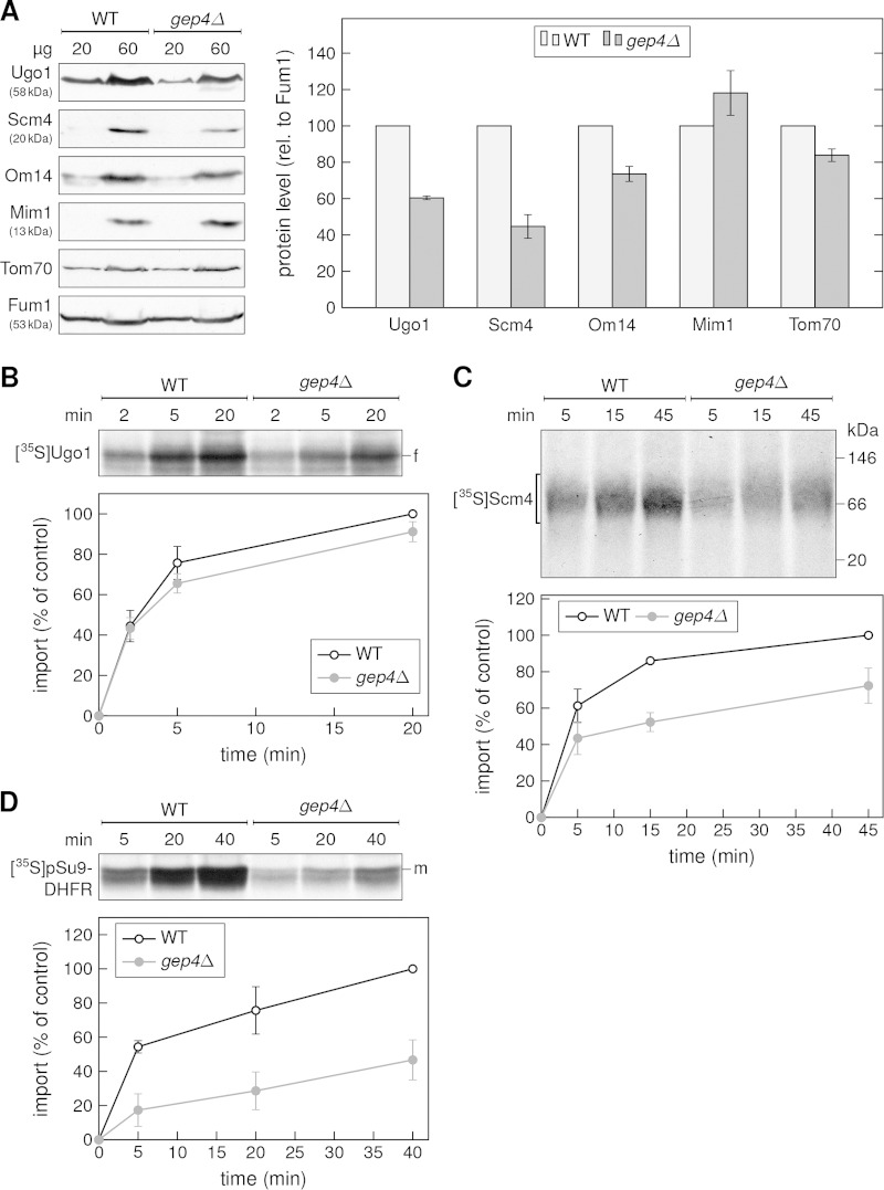
A special group of mitochondrial outer membrane (MOM) proteins spans the membrane several times via multiple helical segments. Such multispan proteins are synthesized on cytosolic ribosomes before their targeting to mitochondria and insertion into the MOM. Previous work recognized the import receptor Tom70 and the mitochondrial import (MIM) complex, both residents of the MOM, as required for optimal biogenesis of these proteins. However, their involvement is not sufficient to explain either the entire import pathway or its regulation. To identify additional factors that are involved in the biogenesis of MOM multispan proteins, we performed complementary high-throughput visual and growth screens in Saccharomyces cerevisiae. Cardiolipin (CL) synthase (Crd1) appeared as a candidate in both screens. Our results indeed demonstrate lower steady-state levels of the multispan proteins Ugo1, Scm4, and Om14 in mitochondria from crd1Δ cells. Importantly, MOM single-span proteins were not affected by this mutation. Furthermore, organelles lacking Crd1 had a lower in vitro capacity to import newly synthesized Ugo1 and Scm4 molecules. Crd1, which is located in the mitochondrial inner membrane, condenses phosphatidylglycerol together with CDP-diacylglycerol to obtain de novo synthesized CL molecules. Hence, our findings suggest that CL is an important component in the biogenesis of MOM multispan proteins.


Figures

FIG 1
FIG 1
Establishment of an in vivo visual assay to monitor the biogenesis of Om14. (A) The GFP-Om14 query strain expressing mitochondrial Aco2-Cherry was subjected to fluorescence microscopy. DIC, differential inference contrast. (B) Subcellular fractionation of the GFP-Om14 query strain. Equal amounts of whole-cell lysate (WCL) and of fractions corresponding to cytosol (cyt), ER, and mitochondria (mito) were analyzed by SDS-PAGE and immunodecoration. The indicated antibodies against marker proteins for mitochondria (Om14 and Tom70), cytosol/nucleus (Bmh1), and ER (Erv2) were used. (C) Carbonate extraction (CE) of mitochondria isolated from either the wild-type strain or the GFP-Om14 query strain was performed, and samples were separated to membrane-embedded proteins in the pellet fraction (P) and soluble proteins in the supernatant (SN). Where indicated (lanes 4 and 8), additional mitochondrial samples were treated with 20 μg/ml proteinase K (PK). All samples were analyzed by SDS-PAGE and immunodecoration. Porin, MOM protein resistant to PK; Tom20, MOM protein sensitive to PK; aconitase, matrix protein resistant to PK. (D) Fluorescence microscopy of representative deletion strains expressing GFP-Om14. (E) Cellular functions of proteins identified as hits by the visual screen. Functions have been assigned according to the Saccharomyces Genome Database.
FIG 2
FIG 2
GFP-Om14 and GFP-Ugo1 have an altered appearance in crd1Δ and gim2Δ deletion strains. Strains with BDH2 (as a control), CRD1, or GIM2 deleted, expressing either GFP-Om14 (upper panels) or GFP-Ugo1 (lower panels), were observed by fluorescence microscopy. Selected regions from each image were enlarged and are shown separately.
FIG 3
FIG 3
Growth assay to monitor the biogenesis of Om14. (A) Schematic representation of the various fusion proteins used. The expected potential of these proteins to support growth on synthetic medium without uracil is indicated at the bottom. (B) Yeast cells expressing the indicated constructs were analyzed at 30°C by drop-dilution assay on synthetic medium lacking either leucine only (SD-Leu) or leucine and uracil (SD-Leu-Ura). (C) Subcellular fractionation of the Ura3-HA-Om14-SL17 query strain. Equal amounts of whole-cell lysate (WCL) and of fractions corresponding to cytosol (cyt), ER, and mitochondria (mito) were analyzed as described in the Fig. 1B legend. (D) Mitochondria isolated from either the WT strain or the Ura3-HA-Om14-SL17 query strain were treated and analyzed as described in the Fig. 1C legend. (E) crd1Δ and cmk2Δ (as a control) cells expressing Ura3-HA-Om14-SL17 were grown on galactose-containing liquid medium in the presence (SGal comp.) or absence (SGal-Ura) of uracil. The optical density at 600 nm (OD600) of the cultures was measured and is depicted as a function of incubation time.
FIG 4
FIG 4
The absence of cardiolipin affects the steady-state levels of multispan proteins. (A) Steady-state levels of GFP-Om14 are decreased in crd1Δ mitochondria. (Left panel) Crude mitochondria from wild-type (WT) and crd1Δ yeast strains were isolated and analyzed by SDS-PAGE and immunodecoration with the indicated antibodies. (Right panel) The bands resulting from three independent experiments were quantified and normalized to the level of Tom20, and the protein levels in the WT strain were set to 100%. Error bars represent standard deviations (n = 3). rel., relative. (B) Steady-state levels of Ugo1 and Scm4 are decreased in crd1Δ organelles. Mitochondria were isolated from either WT or crd1Δ cells, and the indicated amounts were analyzed as described for panel A. The resulting bands were quantified and normalized to the level of Yah1, and the protein levels in the WT strain were set to 100% (right panel). Error bars represent standard deviations (n = 6 [3 biological experiments with 2 technical repeats each]). (C) Mitochondria isolated as described for panel B were subjected to blue native PAGE and immunodecoration with antibodies against Mim1 or Tom40. The MIM and the TOM complexes are indicated.
FIG 5
FIG 5
Mitochondria lacking Crd1 have a lower in vitro capacity to import multispan proteins. (A) Radiolabeled molecules of Ugo1-2HA were incubated with mitochondria isolated from either WT or crd1Δ cells for the indicated time periods. After import, mitochondria were treated with trypsin and subjected to SDS-PAGE and autoradiography. The trypsin-protected fragment is indicated (f). (Lower panel) Bands representing this fragment were quantified, and the intensity of the band upon import into control mitochondria for 20 min was set as 100%. Data represent averages of the results of three independent experiments. (B) Radiolabeled molecules of Scm4 were incubated with isolated mitochondria as described for panel A. After import, mitochondria were solubilized by the use of digitonin and subjected to blue native PAGE and autoradiography. The Scm4-containing band is indicated. (Lower panel) Bands were quantified, and the intensity of the band upon import into control mitochondria for 45 min was set as 100%. Data represent averages of the results of three independent experiments. (C) Radiolabeled molecules of pSu9-DHFR were incubated with isolated mitochondria as described for panel A. After import, mitochondria were treated with proteinase K (PK) and subjected to SDS-PAGE and autoradiography. The PK-protected mature form is indicated (m). (Lower panel) Bands representing the mature form were quantified, and the intensity of the band upon import into control mitochondria for 40 min was set as 100%. Data represent averages of the results of three independent experiments.
FIG 6
FIG 6
Deletion of Gep4 causes a reduction in the biogenesis of multispan proteins. (A) Mitochondria were isolated from either WT or gep4Δ cells, and the indicated amounts were analyzed as described in the Fig. 4B legend. The resulting bands were quantified and normalized to the level of fumarase (Fum1), and the protein levels in the WT strain were set to 100% (right panel). (B to D) The indicated radiolabeled proteins were incubated with isolated organelles and further analyzed as described in the Fig. 5 legend.

References

    1. Dukanovic J, Rapaport D. 2011. Multiple pathways in the integration of proteins into the mitochondrial outer membrane. Biochim Biophys Acta 1808:971–980. doi:10.1016/j.bbamem.2010.06.021. - DOI - PubMed
    1. Fritz S, Rapaport D, Klanner E, Neupert W, Westermann B. 2001. Connection of the mitochondrial outer and inner membranes by Fzo1 is critical for organellar fusion. J Cell Biol 152:683–692. doi:10.1083/jcb.152.4.683. - DOI - PMC - PubMed
    1. Otera H, Taira Y, Horie C, Suzuki Y, Suzuki H, Setoguchi K, Kato H, Oka T, Mihara K. 2007. A novel insertion pathway of mitochondrial outer membrane proteins with multiple transmembrane segments. J Cell Biol 179:1355–1363. doi:10.1083/jcb.200702143. - DOI - PMC - PubMed
    1. Becker T, Wenz LS, Kruger V, Lehmann W, Muller JM, Goroncy L, Zufall N, Lithgow T, Guiard B, Chacinska A, Wagner R, Meisinger C, Pfanner N. 2011. The mitochondrial import protein Mim1 promotes biogenesis of multispanning outer membrane proteins. J Cell Biol 194:387–395. doi:10.1083/jcb.201102044. - DOI - PMC - PubMed
    1. Papic D, Krumpe K, Dukanovic J, Dimmer KS, Rapaport D. 2011. Multispan mitochondrial outer membrane protein Ugo1 follows a unique Mim1-dependent import pathway. J Cell Biol 194:397–405. doi:10.1083/jcb.201102041. - DOI - PMC - PubMed

Publication types

MeSH terms

Substances