. 2015 Jun 29;59(2):2499.
doi: 10.4081/ejh.2015.2499.

WT1 expression in the human fetus during development

Affiliations

WT1 expression in the human fetus during development

R Ambu et al. Eur J Histochem. .

Abstract

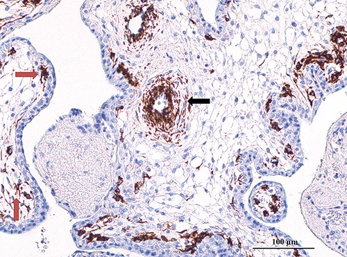
Wilms' Tumor 1 (WT1) is a transcription factor involved in the development of the urogenital system. The purpose of this study was to analyze the immunoreactivity for WT1 protein in different tissues and organs in human fetuses in early phases of gestation. To this end, samples from multiple organs were obtained from 4 human fetuses, ranging from 7 up to 12 weeks of gestation. Each sample was formalin-fixed, paraffin embedded and immunostained for WT1. Our data show that WT1 is involved in development of multiple human organs in a more vast series of cells types than previously reported. Immunostaining for WT1 was characterized by a predominant cytoplasmic reactivity in the vast majority of cell types. Mesenchimal progenitors in the fetal lung, ductal plate progenitors in fetal liver, cap mesenchimal cells in the developing kidney, fetal zone cells in adrenal glands, atrial and ventricular cardiomyocytes in the fetal heart, radial glial cells in the fetal cerebral cortex and skeletal muscle cell precursors showed the highest levels of WT1 immunoreactivity. Future studies will be needed to detect differences in the expression of WT1 in various organs at different gestational ages, in order to better evaluate the role of WT1 in cell proliferation and differentiation during intrauterine human development.


Conflict of interest statement

Conflict of interest: the authors declare that no conflict of interest exist.

Figures

Figure 1.
Figure 1.
WT1 immunostaining in placenta. WT1 immunoreactivity was restricted to cytoplasm of muscular cells in the arterial wall (black arrow) and star-shaped cells in close relationship with the cytotrophoblast (red arrows).
Figure 2.
Figure 2.
WT1 immunostaining in fetal gut. WT1 was detected A) in the cytoplasm of lamina propria of the mucosa and in the fibrovascular axis of villi (black arrows); B) in the submucosa (red arrow).
Figure 3.
Figure 3.
WT1 immunostaining in fetal kidney. WT1 immunoreactivity was detected A) in the developing glomeruli (black arrows) and in the nephrogenic zone located under the renal capsule (red arrow); B) in the cytoplasm of interstitial cells surrounding tubular structures (black arrow); C) in the cytoplasm and nuclei of podocyte precursors (black arrows) and along the developing basal membranes inside glomeruli (red arrow); and D) in the cytoplasm of Cap mesenchimal cells (black arrows) and in spindle cells located in the renal interstitium (red arrow).
Figure 4.
Figure 4.
WT1 immunostaining in fetal adrenal gland. WT1 immunoreactivity was located in the cytoplasm of cells of the fetal zone and in the adrenal capsule, with some strongly reactive capsular cells intermingled with non-reactive cells (F, fetal zone; D, definitive zone; C, adrenal capsule).
Figure 5.
Figure 5.
WT1 immunostaining in fetal lung. WT1 was expressed in cytoplasm of mesenchymal progenitors surrounding branching epithelial structures (black arrows) and in small vessels inside the pulmonary scarcely differentiated mesenchyme (red arrow).
Figure 6.
Figure 6.
WT1 immunostaining in fetal heart. Atrial cardiomyocytes (black arrow) showed higher cytoplasmic levels of WT1 immunostaining as compared to those observed in both ventricles (red arrow).
Figure 7.
Figure 7.
WT1 immunostaining in fetal liver. WT1 cytoplasmic immunoreactivity was observed in the of mesenchymal cells of the developing portal tracts (black arrows) and developing biliary structures (red arrows), including remnants of the ductal plate (green arrow).
Figure 8.
Figure 8.
WT1 immunostaining in fetal central nervous system. WT1 was detected A) in the cytoplasm of Radial glia and in the axons localized in the cerebral cortex (VZ, ventricular zone; MZ, marginal zone) and B) in the cytoplasm of Radial Glia of the spinal cord.
Figure 9.
Figure 9.
WT1 immunostaining in fetal arts. WT1 cytoplasmic immunoreactivity were observed A) in developing skeletal muscles and B) in the progenitor cells of the developing derma (black arrow).

References

    1. Call KM, Glaser T, Ito CY, Buckler AJ, Pelletier J, Haber DA, et al. Isolation and characterization of a zinc finger polypeptide gene at the human chromosome 11 Wilms’ tumor locus. Cell. 1990;60:509-520. - PubMed
    1. Rauscher FJ 3rd, Morris JF, Tournay OE, Cook DM, Curran T. Binding of the Wilms’ tumor locus zinc finger protein to the EGR-1 consensus sequence. Science 1990;250:1259-62. - PubMed
    1. Hamilton TB, Barilla KC, Romaniuk PJ. High affinity binding sites for the Wilms’ tumour suppressor protein WT1. Nucleic Acids Res 1995;23:277-84. - PMC - PubMed
    1. Hastie ND. The genetics of Wilms’ tumor - a case of disrupted development. Annu Rev Genet 1994;28:523-558. - PubMed
    1. Haber DA, Sohn RL, Buckler AJ, Pelletier J, Call KM, Housman DE. Alternative splicing and genomic structure of the Wilms tumor gene WT1. Proc Natl Acad Sci USA 1991;88:9618-22. - PMC - PubMed