What Safe Zone? The Vast Majority of Dislocated THAs Are Within the Lewinnek Safe Zone for Acetabular Component Position
- PMID: 26150264
- PMCID: PMC4709312
- DOI: 10.1007/s11999-015-4432-5
What Safe Zone? The Vast Majority of Dislocated THAs Are Within the Lewinnek Safe Zone for Acetabular Component Position
Abstract
Background: Numerous factors influence total hip arthroplasty (THA) stability including surgical approach and soft tissue tension, patient compliance, and component position. One long-held tenet regarding component position is that cup inclination and anteversion of 40° ± 10° and 15° ± 10°, respectively, represent a "safe zone" as defined by Lewinnek that minimizes dislocation after primary THA; however, it is clear that components positioned in this zone can and do dislocate.
Questions/purposes: We sought to determine if these classic radiographic targets for cup inclination and anteversion accurately predicted a safe zone limiting dislocation in a contemporary THA practice.
Methods: From a cohort of 9784 primary THAs performed between 2003 and 2012 at one institution, we retrospectively identified 206 THAs (2%) that subsequently dislocated. Radiographic parameters including inclination, anteversion, center of rotation, and limb length discrepancy were analyzed. Mean followup was 27 months (range, 0-133 months).
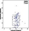
Results: The majority (58% [120 of 206]) of dislocated THAs had a socket within the Lewinnek safe zone. Mean cup inclination was 44° ± 8° with 84% within the safe zone for inclination. Mean anteversion was 15° ± 9° with 69% within the safe zone for anteversion. Sixty-five percent of dislocated THAs that were performed through a posterior approach had an acetabular component within the combined acetabular safe zones, whereas this was true for only 33% performed through an anterolateral approach. An acetabular component performed through a posterior approach was three times as likely to be within the combined acetabular safe zones (odds ratio [OR], 1.3; 95% confidence interval [CI], 1.1-1.6) than after an anterolateral approach (OR, 0.4; 95% CI, 0.2-0.7; p < 0.0001). In contrast, acetabular components performed through a posterior approach (OR, 1.6; 95% CI, 1.2-1.9) had an increased risk of dislocation compared with those performed through an anterolateral approach (OR, 0.8; 95% CI, 0.7-0.9; p < 0.0001).
Conclusions: The historical target values for cup inclination and anteversion may be useful but should not be considered a safe zone given that the majority of these contemporary THAs that dislocated were within those target values. Stability is likely multifactorial; the ideal cup position for some patients may lie outside the Lewinnek safe zone and more advanced analysis is required to identify the right target in that subgroup.
Level of evidence: Level III, therapeutic study.
Figures


References
-
- Berry DJ. Unstable total hip arthroplasty: detailed overview. Instr Course Lect. 2001;50:265–274. - PubMed
MeSH terms
LinkOut - more resources
Full Text Sources
Other Literature Sources
Medical
Research Materials

