Review
. 2015 Oct;21(5):460-74.
doi: 10.1177/1073858415592377. Epub 2015 Jul 6.

Targeting Neuronal Networks with Combined Drug and Stimulation Paradigms Guided by Neuroimaging to Treat Brain Disorders

Affiliations
Review

Targeting Neuronal Networks with Combined Drug and Stimulation Paradigms Guided by Neuroimaging to Treat Brain Disorders

Carl L Faingold et al. Neuroscientist. 2015 Oct.

Abstract

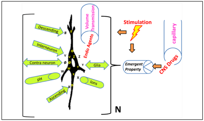
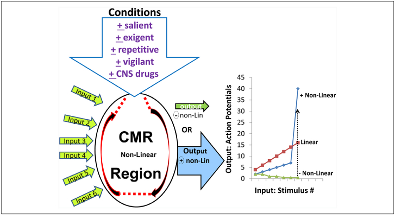
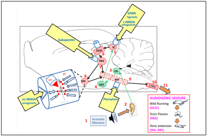
Improved therapy of brain disorders can be achieved by focusing on neuronal networks, utilizing combined pharmacological and stimulation paradigms guided by neuroimaging. Neuronal networks that mediate normal brain functions, such as hearing, interact with other networks, which is important but commonly neglected. Network interaction changes often underlie brain disorders, including epilepsy. "Conditional multireceptive" (CMR) brain areas (e.g., brainstem reticular formation and amygdala) are critical in mediating neuroplastic changes that facilitate network interactions. CMR neurons receive multiple inputs but exhibit extensive response variability due to milieu and behavioral state changes and are exquisitely sensitive to agents that increase or inhibit GABA-mediated inhibition. Enhanced CMR neuronal responsiveness leads to expression of emergent properties--nonlinear events--resulting from network self-organization. Determining brain disorder mechanisms requires animals that model behaviors and neuroanatomical substrates of human disorders identified by neuroimaging. However, not all sites activated during network operation are requisite for that operation. Other active sites are ancillary, because their blockade does not alter network function. Requisite network sites exhibit emergent properties that are critical targets for pharmacological and stimulation therapies. Improved treatment of brain disorders should involve combined pharmacological and stimulation therapies, guided by neuroimaging, to correct network malfunctions by targeting specific network neurons.

Keywords: CNS disorders; CNS pharmacology; complexity; electrical stimulation; emergent property; neuroimaging; neuronal network; pain; seizures; startle.


Conflict of interest statement

Declaration of Conflicting Interests

The authors declare no potential conflict of interest with respect to the research, authorship, and/or publication of this article.

Figures

Figure 1.
Figure 1.
Numerous influences converge on principal neurons leading to emergent properties. This is an idealized diagram of a principal neuron in a specific nucleus within a neuronal network in the brain of an awake, behaving organism that illustrates many of the influences that affect this “class” of neurons (N) in this nucleus. The neurons possess certain specific intrinsic properties (Φ), such as the propensity to exhibit burst firing or pacemaker activity. These cells also possess specific receptors (#) (metabotropic or ionotropic) onto which descending projections release a specific neuroactive substance. The neurons also possess ligand-gated receptors (V) (e.g., glutamate) (which have specific receptor subunits) onto which interneurons synaptically release a specific neuroactive substance. Projection neurons across the midline for bilaterally connected structures releasea neuroactive substance ø, which binds to its specific receptors on contralateral neurons. The principal neurons also possess the property of pH sensitivity (W). Ascending input from neuronsin nuclei in the network also release a neuroactive substance (¤), which binds to its specific receptors. The neurons possess voltage-gated ion channels (X) (e.g., K+ channels) at which local ions can act. The neurons receive input from local glial cells, which release a neuroactive substance (Y) (e.g., adenosine) that acts on specific receptors for this substance. Endogenous (Endo) neuroactive agents (Z) carried via volume transmission from nearby (spillover) or distant sites via the extracellular fluid and cerebrospinal fluid to the neurons also affect the properties of these neurons. An example would be extrasynaptic GABAA receptors that respond to the low levels of “ambient” GABA in the extracellular fluid. Finally, when an exogenous agent, such asa CNS drug or centrally acting toxin, is administered it is released via the brain blood vessels among other vectors to exert its effects on the emergent property of these principal cells to exert its effect on network function. The therapeutic effect of many CNS drugs is postulated to be mediated by a selective action on the principal cells in a specific nucleus in the neuronal network for the disorder to produce the desired effect. This selectivity is seen primarily when therapeutic doses are given. In addition, electrical, magnetic, or optogenetic neurostimulation can modulate the network and affect the emergent property. The summation of all these influences determines whether the emergent property is expressed and the actual nature of that property and its relative sensitivity in that group of neurons. This emergent property is postulated to be expressed uniquely in this network nucleus and causes this class of neurons to respond with unique sensitivityto a specific exogenous substance. Note, those influences can also be modified by brain state changes (such as sleep), whichcan significantly alter the emergent property, based on changesin milieu of the principal neuron as well as external stimulation. (Modified from Faingold 2014b, with permission.)
Figure 2.
Figure 2.
Neuroimaging reveals consciousness networks. Blood oxygen level dependent (BOLD) fMRI during seizurein a rodent model of complex partial seizures demonstrates coordinated cortical and subcortical changes. Seizures are induced by hippocampal stimulation (yellow arrowheads indicate electrodes), which leads to seizure activity and an increased BOLD signal (warm colors) in the hippocampus (HC), septal nuclei, and anterior hypothalamus (Ant Hyp). This causes cortical slow oscillations (not shown) and a decreased BOLD signal (cool colors) in the orbital frontal cortex (LO/VO), as well as in subcortical arousal areas such as the intralaminar thalamic centrolateral nucleus (CL) and midbrain tegmentum (MT). Limbic seizures of this kind are associated with behavioral arrest and decreased responsiveness in both animal models and human patients with temporal lobe epilepsy. (Motelow and others 2015, with permission.)
Figure 3.
Figure 3.
Input-output relationships of a prototypical conditional multireceptive (CMR) brain region, which is capable of a significant degree of self-organization, as shown by the paired semicircular arrows. This diagram illustrates the numerous inputs that the neurons in this CMR region receive from primary networks and other CMR networks. The output of neuronsin CMR nuclei is highly dependent on the conditions that the animal is experiencing, including salient, exigent, and repetitive conditions and can also be governed by the animal’s state of vigilance as well as centrally acting pharmacological agents. The output of CMR regions is subject to nonlinearity wherein CMR neuronal responsiveness to any input can change dramatically from nonresponsiveness to hyperresponsiveness, depending on stimulus parameter type, strength, and repetition rate, as illustrated in the graph on the right. Under nonexigent (resting) conditions many CMR neurons exhibit negative (−) nonlinearity with minimal responsiveness or even nonresponsiveness to the input, as seen in neurons that exhibit response habituation. The various exigent conditions can cause CMR neurons to exhibit hyperresponsiveness and exhibit positive (+) nonlinearity, which can result in massive output, activating the self-organization characteristics of the CMR network and result in emergent properties of that network, which range, for example, from startle responses to generalized seizures. CMR neurons that exhibit negative nonlinearity under resting conditions can convert to positive nonlinearity in response to exigent changes in the animal’s behavioral state (dotted line). The nonlinear output characteristics of CMR brain regions contrast with the relatively linear responses seen in primary networks, such as sensory systems. (Based on Faingold and others 2014b, with permission.)
Figure 4.
Figure 4.
Changes in conditional multireceptive (CMR) neuronal responses induced by CNS depressant drugs. Neuronal responses in the ventrolateral periaqueductal gray (PAG) and lateral amygdala (LA) are depressed by low doses of depressant drugs. Low doses of barbiturates (which enhance GABAA receptor activity) or ketamine (uncompetitive NMDA receptor antagonist) reduce CMR extracellular action potential responses to external stimuli. Panel A shows the mean (± SEM) reduction of both spontaneous (Spont.) and thermally evoked PAG neuronal firing by pentobarbital (N = 18 neurons) 15 minutes after systemic administration with recovery by 30 minutes. Panel B shows examples of rate meter histogram analysis of a PAG spontaneous and evoked neuronal firing in response to a single 30-second (bracket) noxious thermal stimulation (53°C) before (Control column), 15 minutes after pentobarbital (15 mg/kg i.p.) treatment (Drug column), and subsequent recovery (at 30 minutes) (Recovery column). Prior to pentobarbital the majority (18/35) of PAG neurons increased firing in response to the thermal stimulus in this study. (Note 10 mg/kg of pentobarbital had no significant effect on PAG neuronal firing, whereas 20 mg/kg produced greater depression of PAG firing than the 15 mg dose.) The action potential above the top histogram represents the waveform of the neuron being analyzed. The onset and duration of the thermal stimulus is illustrated by the bracket. *Significance at P < 0.01 (repeated-measures ANOVA). Panel C shows a representative example of the poststimulus time histogram (PSTH) analysis of the action of an uncompetitive NMDA antagonist (ketamine) to reversibly block the sensory responsiveness of LA neurons. In the control column the LA neuron exhibited an onset response to the auditory stimulus before ketamine treatment. LA neuronal firing was significantly reduced (P < .01, paired t-test) and almost completely suppressed 15 minutes after ketamine (30 mg/kg i.p.) treatment (drug column). Four hours after ketamine treatment, the LA neuronal response was comparable tothat prior to ketamine treatment (recovery column). The inset the control column in line C shows an example of the digital oscilloscope tracings for the PSTH. Action potential amplitude in C: 300 μV. N = number of action potentials in the PSTH. Treatment was given in unanesthetized awake, behaving rats with microwire recording electrode. (Acoustic stimulus parameters: 12 kHz tone burst, 100 ms duration, 5 ms rise-fall, 100 dB SPL, 0.5 Hz rate) (PSTH parameters 50 stimulus presentations, 1 ms bin width). All data were taken from awake, behaving rats. The data in line C is representative of the change in the mean number of action potentials/PSTH (control mean = 182.1 ± 42.0 [SEM] vs. 9.6 ± 2.0 drug mean). Note: Both drug doses were less than 30% of the anesthetic dose. (A, B: modified from Faingold and others 2014b; C: modified from Faingold 2014b).
Figure 5.
Figure 5.
Changes in extracellular action potential firing of conditional multireceptive (CMR) neurons in several brain regionsin response to sensory stimuli induced by administration of GABAA receptor antagonists (in subconvulsant doses). In predrug conditions (control column of the poststimulus histograms or PSTHs) presentations of electrical stimuli to the sciatic nervein line B to a mesencephalic reticular formation (MRF) neuron, line C amygdala (AMG) neuron response to auditory stimuli, line D response of a pericruciate cortex (CTX) neuron to visual stimuli and response of a MRF neuron to electrical stimulus within the brain to the inferior colliculus (IC) in (line E). Although spontaneous firing was observed, very little evidence of responsiveness to the stimuli is detectable, as shown by the absence of clear time-locked response peaks to the stimuli in the PSTHs (control column). After administration of a GABAA receptor antagonist these CMR neurons exhibit major responsiveness increases, which includes the induction of a very striking time-locked peak of responsiveness to each stimulus in each PSTH (drug column). These peaks indicate that the drug had induced extensive responsiveness of the CMR neurons to these stimuli. The bar graphs in A compare changes in the PSTH peak period and the rest of the recorded period for the PSTHs in line B. The peak (20–55 ms period after stimulus onset) shows a much greater percent increase as compared to the rest of the 400 ms sampling period as shown by the bar graphs for the nonpeak versus peak time points shown above each PSTH. Several of these CMR neurons showed response enhancement to more than one stimulus modality (not shown), indicating the multimodality responsiveness common in CMR neurons. This neuroplastic effect is short-term, since the firing patterns of these neurons recover to unresponsiveness with time (right column). Specific GABAA receptor antagonists and doses used are as follows: line B, pentylenetetrazol, 15 mg/kg; line C, bemegride, 3.6 mg/kg; line D, bicuculline, 0.03 mg/kg; line E, pentylenetetrazol, 5 mg/kg. Recovery times ~20 minutes after i.v. drug administration. Stimulus parameters: 0.5 Hz stimulus rate, 50 stimulus presentations (100 μs single bipolar pulses in B and E, 95 dB SPL clicks in C, and 18.5 lux visual stimulus [strobe] in D). PSTHs: bin width = 1 ms, scan length= 400 ms. Note the histograms in line D are peristimulus time histograms. N = number of action potentials per PSTH. This effect was seen in 88% of more than 700 bulbar and midbrain reticular formation neurons, 57% of more than 100 AMG, and 67% of more than 100 CTX neurons in unanesthetized cat and rat and was also seen with direct (iontophoretic) application of drug. (A, B, E: modified from Faingold and Feng 2014; C: modified from Faingold 2014b; D: modified from Faingold and others 1983.)
Figure 6.
Figure 6.
Diagram of the neuronal network for audiogenic seizures (AGSz) with emergent properties (indicated by cylinders) in each requisite site at which systemically administered drugs that block these seizures may act at therapeutic doses. The network is organized as a hierarchy beginning with the acoustic stimulus (1) input into the auditory pathway (2–4), including neurons in these nuclei (up to the level of the inferior colliculus, IC) (5), whichis the consensus seizure-initiating site. The IC projects to the brainstem locomotion network nuclei, including the deep layersof superior colliculus (DLSC) (6), projecting to the periaqueductal gray (PAG) (7) and substantia nigra reticulata (SNr) (8) and brainstem reticular formation (BRF) (9), which project to the spinal cord (10). The hierarchical activation of each requisite network nucleus produces the sequential behaviors of AGSz (wild running followed by tonic flexion and tonic extension) (11). (The critical structure that initiates each behavior is shown below the behavior.) Neurons in the BRF and SNr are the only regions that are active during the postictal behavioral depression that follows the tonic extension behavior. Therapeutic doses of several drugs with anticonvulsant properties act to inhibit neurons in the IC, including competitive (c-) NMDA antagonists, such as 2-amino-7-phosphonoheptanoate and GABA uptake inhibitor, tiagabine, as well as ethanol. Therapeutic doses of other drugs that are effectively anticonvulsant exert no effect on IC neurons, including an uncompetitive (uc-) NMDA antagonist (MK-801), gabapentin, lamotrigine, and phenytoin. Other effective anticonvulsant drugs act to reduce PAG neuronal firing such as gabapentin. Therapeutic doses of phenytoin selectively act to inhibit neurons in the BRFof the pons. The SNr is the target of MK-801, but the effect is to increase neuronal firing. The emergent property of each nucleus is seen as a confluence of influences onto the neurons in each nucleus, including neuroactive substances (red squares) released onto the neurons via synaptic transmission (ST) and from the blood vessels, cerebrospinal and extracellular fluids via volume transmission (VT), as shown in the expanded diagram of an emergent property on the left of the network. Systemically administered drugs reach each site via VT. Direct stimulation of any site within the network, either chemically or electrically, can affect seizure susceptibility and may modify the emergent properties in the affected site. (CN = cochlear nucleus; SOC = superior olivary complex, which are auditory structures important for input to the IC.) Brain regions in the auditory pathway rostral to the IC were not requisite to seizure induction, and in fact, no regions rostral to the level of the midbrain are requisite to these seizures. It should be noted that neurons in the lateral amygdala are affected by these seizures, but blockade of this region does not block seizures, rendering the amygdala as an ancillary site.

Similar articles

Cited by

References

    1. Agnati LF, Genedani S, Spano P, Guidolin D, Fuxe K. 2014. Volume transmission and the Russian-doll organization of brain cell networks: aspects of their integrative actions In: Faingold C, Blumenfeld H, editors. Neuronal networks in brain function, CNS disorders, and therapeutics. San Diego, CA: Academic Press; p. 103–19.
    1. Allen L, Mehta S, McClure JA, Teasell R. 2012. Therapeutic interventions for aphasia initiated more than six months post stroke: a review of the evidence. Top Stroke Rehabil 19(6):523–35. - PubMed
    1. Aupperle RL, Tankersley D, Ravindran LN, Flagan T, Stein NR, Stein MB, and others. 2012. Pregabalin effects on neural response to emotional faces. Front Hum Neurosci 6:42. - PMC - PubMed
    1. Barbarisi M, Pace MC, Passavanti MB, Maisto M, Mazzariello L, Pota V, and others. 2010. Pregabalin and transcutaneous electrical nerve stimulation for postherpetic neuralgia treatment. Clin J Pain 26(7):567–72. - PubMed
    1. Bellani M, Dusi N, Yeh PH, Soares JC, Brambilla P. 2011. The effects of antidepressants on human brain as detected by imaging studies. Focus on major depression. Prog Neuropsychopharmacol Biol Psychiatry 35(7):1544–52. - PubMed

Publication types

LinkOut - more resources