. 2015 Sep 3;43(15):7624-37.
doi: 10.1093/nar/gkv689. Epub 2015 Jul 6.

Structural and functional studies of the Mycobacterium tuberculosis VapBC30 toxin-antitoxin system: implications for the design of novel antimicrobial peptides

Affiliations

Structural and functional studies of the Mycobacterium tuberculosis VapBC30 toxin-antitoxin system: implications for the design of novel antimicrobial peptides

In-Gyun Lee et al. Nucleic Acids Res. .

Abstract

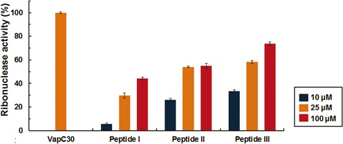
Toxin-antitoxin (TA) systems play important roles in bacterial physiology, such as multidrug tolerance, biofilm formation, and arrest of cellular growth under stress conditions. To develop novel antimicrobial agents against tuberculosis, we focused on VapBC systems, which encompass more than half of TA systems in Mycobacterium tuberculosis. Here, we report that theMycobacterium tuberculosis VapC30 toxin regulates cellular growth through both magnesium and manganese ion-dependent ribonuclease activity and is inhibited by the cognate VapB30 antitoxin. We also determined the 2.7-Å resolution crystal structure of the M. tuberculosis VapBC30 complex, which revealed a novel process of inactivation of the VapC30 toxin via swapped blocking by the VapB30 antitoxin. Our study on M. tuberculosis VapBC30 leads us to design two kinds of VapB30 and VapC30-based novel peptides which successfully disrupt the toxin-antitoxin complex and thus activate the ribonuclease activity of the VapC30 toxin. Our discovery herein possibly paves the way to treat tuberculosis for next generation.


Figures

Figure 1.
Figure 1.
Overall structure of the Mycobacterium tuberculosis VapBC30 complex. (A) Ribbon representation of the VapBC30 heterotetramer of crystal form I, which is nearly identical to the VapBC30 heterotetramer of crystal form II. The models of the VapC30 toxin are colored in orange (chain A) and yellow (chain C), respectively. The models of the VapB30 antitoxin are colored in blue (chain B) and purple (chain D), respectively. The disordered regions (Gly89–Arg92) of the loop connecting α5 and α6 are depicted as dotted lines. The pseudo-2-fold axis is indicated as a black oval. (B) Stereo view of the M. tuberculosis VapBC30 heterodimer formed by chains A (orange) and B (blue) of crystal form I. The α–helices and β–strands of the VapC30 fold are labeled as α1 to α6 and β1 to β4, respectively. An α helix and a 310 helix of the VapB30 fold are labeled as α1 and η1, respectively. The disordered regions (Gly89–Arg92) of the loop connecting α5 and α6 are depicted as dotted lines. (C) A topology diagram of the VapBC30 complex. VapC30 and VapB30 are colored in orange and blue, respectively. The helices and strands are indicated by cylinders and arrows, respectively. (D) The electrostatic surface potential of VapC30 (chain A of crystal form I) is plotted at ±3 kT/e and shown with the ribbon representation of VapB30 (chain B of crystal form I, colored in blue). The active site cavity is indicated by black dotted circle. The electrostatic surface potential was calculated without Mg2+ ion (Please see Supplementary Figure S4A for the electrostatic surface potential calculated with Mg2+ ion). (E) Structures of homologs of M. tuberculosis VapBC30 [Shigella flexneri VapBC (PDB code 3TND); Rickettsia felis VapBC2 (PDB code 3ZVK); M. tuberculosis VapBC15; Neisseria gonorrhoeae FitAB (PDB code 2FE1)]. All structures are drawn in nearly the same orientation as in (B). The models of toxins and antitoxins are colored in orange and blue, respectively.
Figure 2.
Figure 2.
Sequence alignment of PIN-domain family proteins S. flexneri VapC, R. felis VapC2, M. tuberculosis VapC15, Pyrobaculum aerophilum VapC3 and N. gonorrhoeae FitB. Strictly conserved residues and highly conserved residues are indicated by red and yellow colored boxes, respectively. The conserved acidic residues discussed in the text are marked with red asterisks. The residues involved in the binding of proximal and distal antitoxins are marked with green triangles and cyan circles, respectively.
Figure 3.
Figure 3.
Detailed structures of the homodimeric interface of M. tuberculosis VapC30. (A) The homodimeric interface of VapC30 is indicated by a blue dotted rectangle. Each active site of VapC30 is indicated by pink dotted circles. The key residues in the active site are colored in red and also depicted in the figure. The detailed interface of the VapC30 homodimer is shown in Figure 3(B). (B) Details of the homodimeric interface of the VapC30 toxin between chains A and C. Upper: the residues involved in hydrophilic interactions at the homodimeric interface. Hydrogen bonds and salt bridges are shown as black dotted lines. Lower: the residues involved in hydrophobic interactions at the homodimeric interface. Residues participating in hydrophobic interactions are also shown in stick models and presented in the same orientation as in the upper panel. (C) Schematic diagrams of the hydrophilic interactions (upper) and hydrophobic interactions (lower) contributed by residues in the homodimeric interface of VapC30.
Figure 4.
Figure 4.
Detailed active site structures of M. tuberculosis VapC30 and homologs. (A) Close-up view of the M. tuberculosis VapC30 active site (chain A of crystal form II). The bound Mg2+ (Mg1) is shown as a red sphere, and the loop (residues 87–96) connecting α5 and α6 is colored in pink. The positively charged region of the loop (residues 89–92) is disordered in crystal form I. The key residues engaged in the hydrogen bonding network are also shown in the stick models. (B) Superposition of key conserved residues of M. tuberculosis VapC30 (yellow carbons) with M. jannaschii FEN-1 (pink), M. tuberculosis VapC3 (cyan), and M. tuberculosis VapC15 (grey). The Mn2+ ions (from M. jannaschii FEN-1 and M. tuberculosis VapC15) and Mg2+ ions (from M. tuberculosis VapC3 and VapC15) are represented as purple and blue spheres, respectively. Mg1 is represented as a red sphere with its difference electron density map (mFo-Fc; contoured at 1σ).
Figure 5.
Figure 5.
M. tuberculosis VapC30 regulates cellular growth through both Mg2+ and Mn2+-dependent ribonuclease activity. (A) Expression of the VapC30 or VapBC30 complex was induced at hour 0, and cell growth was monitored by measuring the optical density at 600 nm (OD600). Red curves indicate cells expressing the VapC30, and the blue curves indicate cells co-expressing the VapC30 and VapB30. The green curves indicate cells carrying empty pCOLD1 vector and pGro7 plasmids. The data (OD600) represent the average of experiments performed in triplicate, with standard deviations indicated by error bars. (B and C) Fluorescence quenching assay. In this assay system, fluorescence increases when the substrate RNA is cleaved. For the assay, 10 μM VapC30 (or VapBC30), 0.5 M NaCl, 20 mM Tris-HCl buffer at pH 8.0, 40 units of RiboLockTM RNase inhibitor (Thermo Scientific), and various concentrations of (B) MgCl2 or (C) MnCl2 were included in a 50 μl reaction volume and incubated at 37°C. For the assay, possible bound metal ions were removed before the assay (see main text for details). Control contained 10 mM MgCl2 (or 10 mM MnCl2), 0.5 M NaCl, 50 mM Tris-HCl buffer pH 8.0, and 40 units of RiboLockTM (Thermo scientific) RNase inhibitor. Each experiment was performed in triplicate. (D) M. tuberculosis tRNAfMET (1 μM) was used to analyze the ribonuclease activity of VapC30 (2, 4, and 6 μM, respectively). For the assay, 0.5 M NaCl, 20 mM Tris-HCl buffer at pH 8.0, and 40 units of RiboLockTM RNase inhibitor (Thermo Scientific) and 1 mM MgCl2 were included in a 10 μl reaction volume and incubated at 37°C for 60 minutes. Digested RNA fragments were analyzed by denaturing 15% acrylamide gel electrophoresis in the presence of 8 M urea.
Figure 6.
Figure 6.
Interactions between M. tuberculosis VapC30 and VapB30. (A) Overview of the heterotetrameric complex shown in surface representations. The pink dotted rectangles indicate the views shown in (B), (C) and (D). (B) The interface between VapC30 and its proximal VapB30 is shown for hydrophilic interactions (left) and hydrophobic interactions (right). Residues participating in hydrophilic interactions (left) are shown in the stick models, and hydrogen bonds and salt bridges are shown as black dotted lines. Residues participating in hydrophobic interactions (right) are shown in the stick models. VapC30 and VapB30 are colored as in Figure 1. (C and D) Recognition and inhibition of VapC30 chain A by its distal VapB30 chain D (C) and VapC30 chain C by its distal VapB30 chain B (D). Residues participating in hydrophilic interactions are shown in the stick models, and hydrogen bonds are shown as black dotted lines.
Figure 7.
Figure 7.
Fluorescence quenching assays with 10 μM VapC30 (or VapBC30 complex) and various concentrations of the peptides [10 μM (blue), 25 μM (orange), and 100 μM (red)]. VapBC30 complex and the peptides were incubated at 37°C for 60 min before measuring the fluorescence. Fluorescence (RFU) obtained with the 10 μM VapC30 was taken as 100% and fluorescence (RFU) obtained with the 10 μM VapBC30 complex was taken as 0%. For the assay, 0.5 M NaCl, 20 mM Tris–HCl buffer at pH 8.0, 1 mM MgCl2, and 40 units of RiboLock™ RNase inhibitor (Thermo Scientific) were included in a 50 μl reaction volume. Error bars represent the standard deviation of three replicate reactions.

References

    1. World Health Organization (WHO) Global Tuberculosis Report 2014. World Health Organization; 2014.
    1. Centers for Disease Control (CDC) Nosocomial transmission of multidrug-resistant tuberculosis among HIV-infected persons–Florida and New York, 1988–1991. MMWR. Morbidity Mortality Weekly Rep. 1991;40:585. - PubMed
    1. Gandhi N.R., Moll A., Sturm A.W., Pawinski R., Govender T., Lalloo U., Zeller K., Andrews J., Friedland G. Extensively drug-resistant tuberculosis as a cause of death in patients co-infected with tuberculosis and HIV in a rural area of South Africa. Lancet. 2006;368:1575–1580. - PubMed
    1. Migliori G., De Iaco G., Besozzi G., Centis R., Cirillo D. First tuberculosis cases in Italy resistant to all tested drugs. Eur. Surveill. 2007;12:E070517. - PubMed
    1. Udwadia Z.F., Amale R.A., Ajbani K.K., Rodrigues C. Totally drug-resistant tuberculosis in India. Clin. Infect. Dis. 2012;54:579–581. - PubMed

Publication types

MeSH terms