. 2015 Jul 21;112(29):9052-7.
doi: 10.1073/pnas.1423967112. Epub 2015 Jul 6.

The EBF transcription factor Collier directly promotes Drosophila blood cell progenitor maintenance independently of the niche

Affiliations

The EBF transcription factor Collier directly promotes Drosophila blood cell progenitor maintenance independently of the niche

Billel Benmimoun et al. Proc Natl Acad Sci U S A. .

Abstract

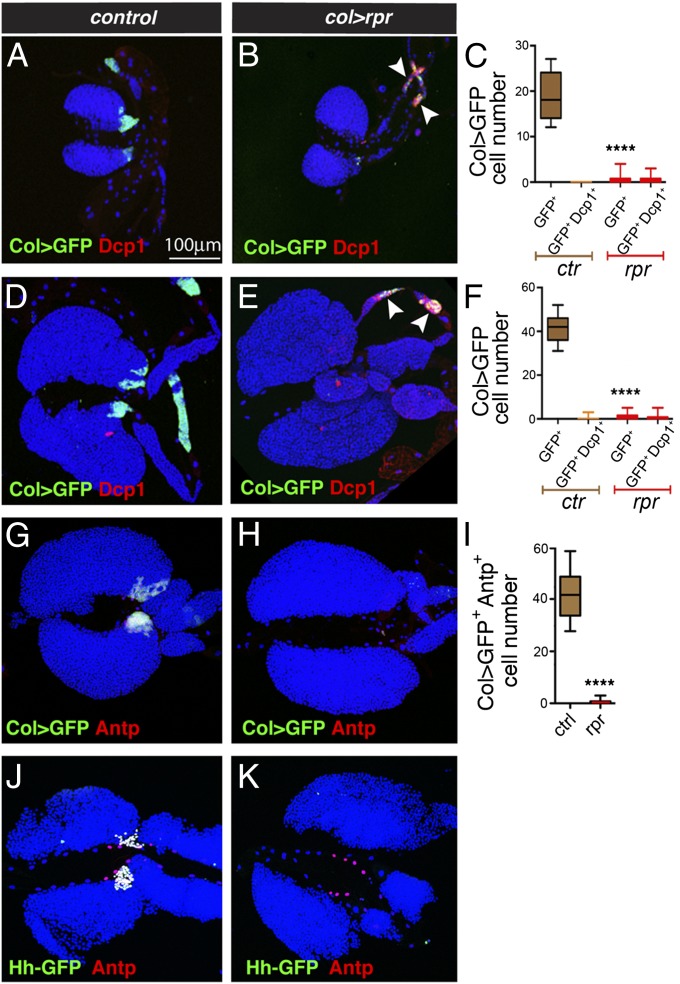
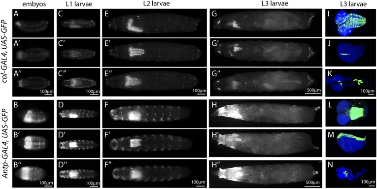
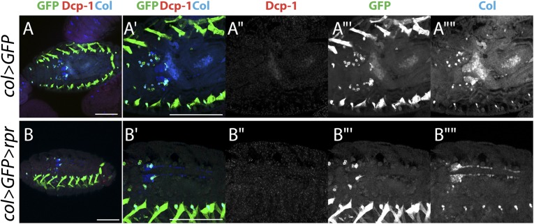
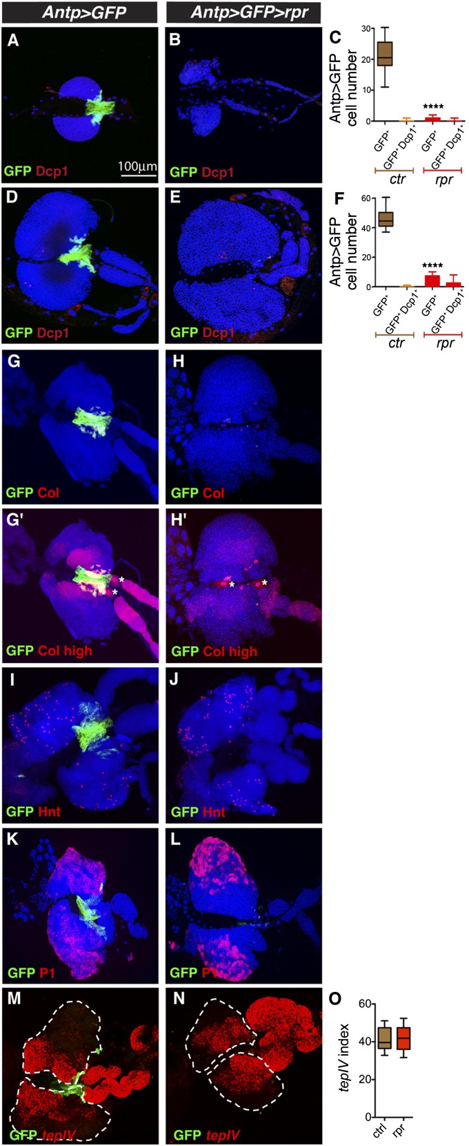
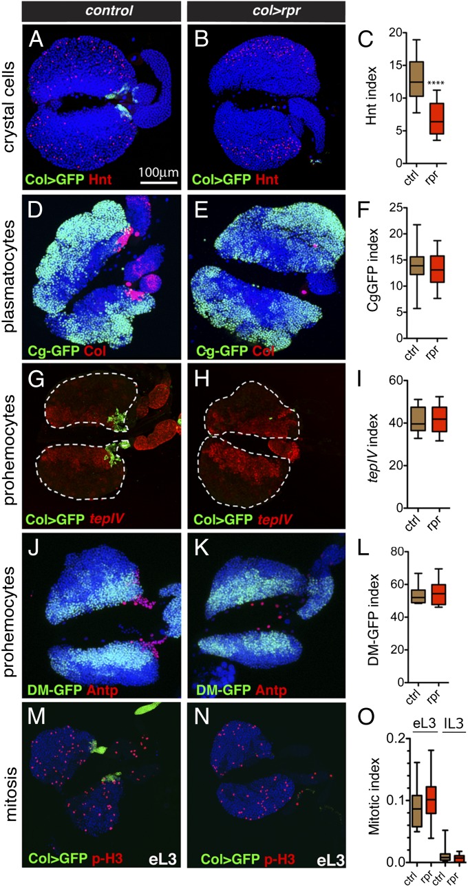
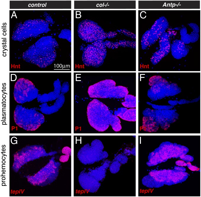
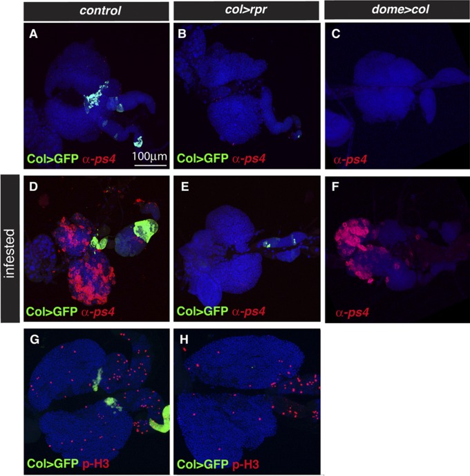
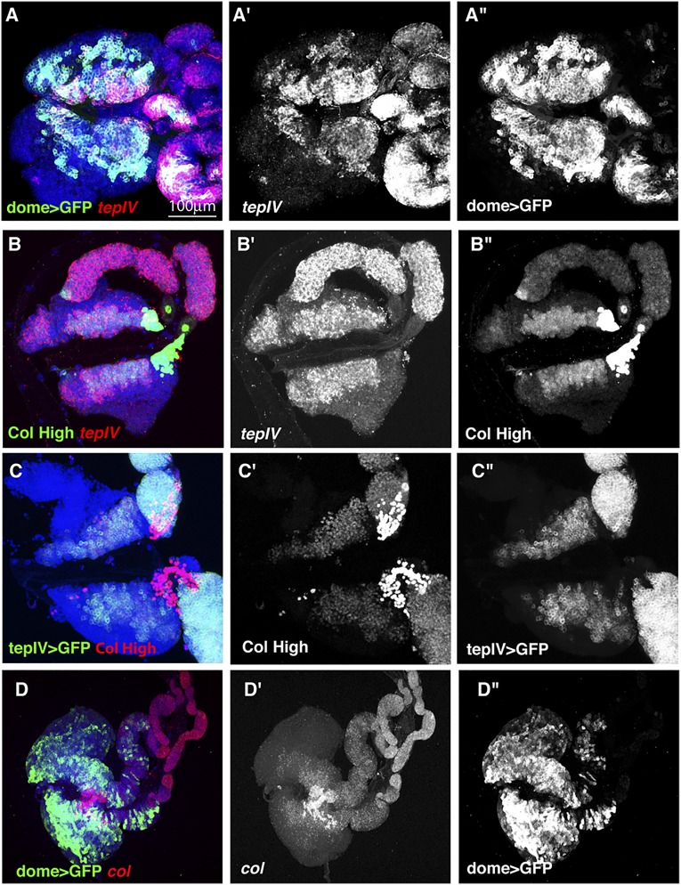
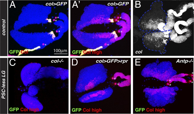
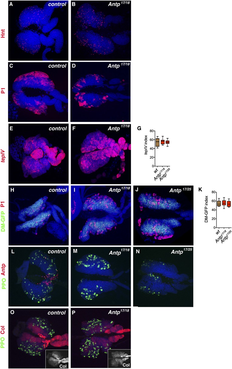
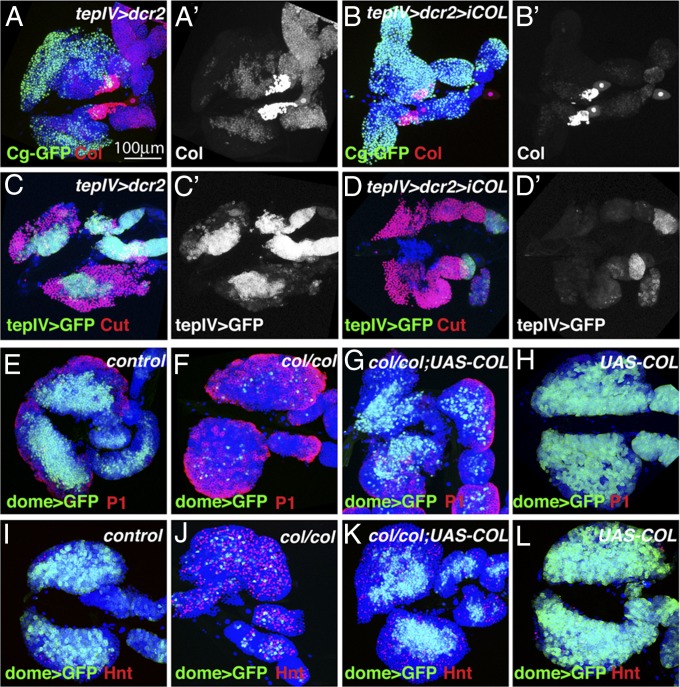
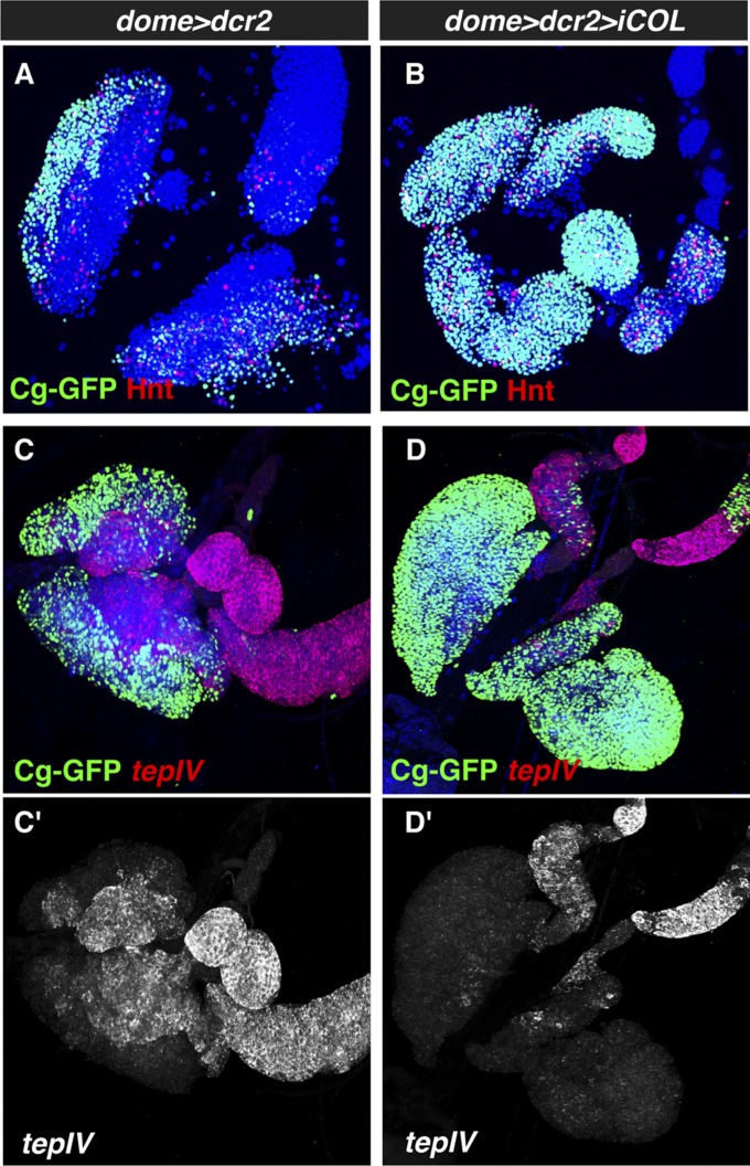
The maintenance of stem or progenitor cell fate relies on intrinsic factors as well as local cues from the cellular microenvironment and systemic signaling. In the lymph gland, an hematopoietic organ in Drosophila larva, a group of cells called the Posterior Signaling Centre (PSC), whose specification depends on the EBF transcription factor Collier (Col) and the HOX factor Antennapedia (Antp), has been proposed to form a niche required to maintain the pool of hematopoietic progenitors (prohemocytes). In contrast with this model, we show here that genetic ablation of the PSC does not cause an increase in blood cell differentiation or a loss of blood cell progenitors. Furthermore, although both col and Antp mutant larvae are devoid of PSC, the massive prohemocyte differentiation observed in col mutant is not phenocopied in Antp mutant. Interestingly, beside its expression in the PSC, Col is also expressed at low levels in prohemocytes and we show that this expression persists in PSC-ablated and Antp mutant larvae. Moreover, targeted knockdown and rescue experiments indicate that Col expression is required in the prohemocytes to prevent their differentiation. Together, our findings show that the PSC is dispensable for blood cell progenitor maintenance and reveal the key role of the conserved transcription factor Col as an intrinsic regulator of hematopoietic progenitor fate.

Keywords: Drosophila; EBF; hematopoiesis; stem cell niche.


Conflict of interest statement

The authors declare no conflict of interest.

Figures

Fig. 1.
Fig. 1.
Rpr expression induces an efficient ablation of the PSC. (A and B) Second and (D and E) third instar larval lymph glands showing GFP and Dcp1 expression in col-GAL4,UAS-mCD8 GFP (A and D, control) or col-GAL4,UAS-mCD8GFP;UAS-rpr (B and E; col > rpr) larvae. Arrowheads: Dcp1+GFP+ cells in the posterior lobes. (C and F) Box-and-whisker representations of the number of GFP+ or GFP+Dcp1+ PSC cells present in control (ctr) or col > rpr (rpr) second (C) and third (F) instar larval lymph glands. (G, H, and I) Antp and GFP expression were used to label PSC cells in col-GAL4,UAS-mCD8-GFP (G) or col-GAL4,UAS-mCD8GFP;UAS-rpr (H) in third instar lymph glands. (I) Box-and-whisker representation of the number of PSC cells (Antp+ GFP+). (J and K) Antp and Hh-GFP expression in HhF4-GFP;col-GAL4 (J) or HhF4-GFP;col-GAL4;UAS-rpr (K) third instar lymph glands. Student’s t test: ****P < 0,0001.
Fig. S1.
Fig. S1.
Expression pattern of col-GAL4 (A, C, E, G, I, J, and K) and Antp-GAL4 (B, D, F, H, L, M, and N). 20xUAS-6xGFP was used as a reporter line. Identical acquisition settings were used for the two drivers. (AH): whole stage 14 embryos (A and B), first (C and D), second (E and F), and third (G and H; spliced images) instar larvae were imaged on an epifluorescence microscope. Lateral (AG), ventral (A’–G’), and dorsal (A”–G”) views of the same individual are shown. (IN) Confocal images of brain (I and L) and wing imaginal discs (J and M), and lymph glands (K and N) from third instar larvae. Nuclei were stained with Topro3.
Fig. S2.
Fig. S2.
Dorsal views of stage 17 embryos showing GFP (green), Dcp1 (red), and Col (blue) expression in col-GAL4,UAS-mCD8GFP (AA””, col > GFP) or col-GAL4,UAS-mCD8GFP;UAS-rpr (BB””, col > rpr) embryos. (A and B) Whole embryos. (A’–A”” and B’–B“”) High-magnification views of the lymph gland region. (Scale bar: 100 μm.)
Fig. S3.
Fig. S3.
Antp-GAL4-driven expression of RPR results in the absence of PSC but does not cause massive hemocyte differentiation. (A, B, D, and E) Second (A and B) and third (D and E) instar larval lymph glands showing GFP and Dcp1 expression in Antp-GAL4,UAS-mCD8GFP;tubGAL80ts (A and D, Antp > GFP) or Antp-GAL4,UAS-mCD8GFP;UAS-rpr, tubGAL80ts (B and E; Antp > GFP > rpr) larvae. (C and F) Quantifications of the number of GFP+ and/or GFP+Dcp1+ cells present in the primary lobes of Antp > GFP (ctr) or Antp > GFP > rpr (rpr) second (C) and third (F) instar larval lymph glands. Box-and-whisker representations. Student’s t test: ****P < 0,0001. (GN) Third instar larval lymph glands. Expression of the PSC/prohemocyte marker Col (G and H: low exposure, G’ and H’: high exposure), the crystal cell marker Hnt (I and J), the plasmatocyte marker P1 (K and L) and the prohemocyte marker tepIV (M and N) in Antp > GFP or Antp > GFP > rpr larvae. Asterisks indicate pericardial cells. (O) Quantification of prohemocyte (tepIV+) population in the primary lobe of Antp > GFP (ctr) and Antp > GFP > rpr (rpr) third instar larvae.
Fig. 2.
Fig. 2.
PSC ablation does not cause prohemocyte loss. Expression of the crystal cell marker Hnt (A and B), the plasmatocyte marker Cg-GFP (D and E), the prohemocyte markers tepIV (G and H) and domeMESO-GFP (DM-GFP; J and K) and the mitotic marker phospho Histone H3 (p-H3; M and N) was assessed in lymph glands from control or col > rpr third instar larvae together with the expression of col > GFP (A, B, G, H, M, and N), Col (D and E) or Antp (J and K). Nuclei were stained with Topro3, except in (G and H) where primary lobes are delineated by dashed white lines. (C, F, I, L, and O) Quantifications of the indicated marker indexes in primary lobes of control (ctrl) and col > rpr (rpr) larvae. Box-and-whisker representations. Student’s t test: ****P < 0,0001. (M, N, and O) eL: early L3 (72 h), lL3: late L3 (116 h).
Fig. 3.
Fig. 3.
Col but not Antp is required for prohemocyte maintenance. (AI) Expression of Hnt (AC), P1 (DF), or tepIV (GI) in wild-type (A, D, and G), col1 (B, E, and H), and Antp17/25 (C, F, and I) third instar larval lymph glands. Nuclei were stained with Topro3.
Fig. S4.
Fig. S4.
(AF) Expression of the lamellocyte marker α-ps4 in early third lymph glands from control (A and D: col-Gal4,UAS-mCD8GFP), col > rpr (D and E: col-Gal4,UAS-mCD8GFP; UAS-rpr), and dome > col (C and F: dome-GAL4;UAS-col) larvae in normal conditions (AC) or following infections by the parasitic wasp L. boulardi (DF). (G and H) Expression of the mitotic marker phospho-Histone H3 (p-H3) in late third instar larval lymph glands from control (G: col-Gal4,UAS-mCD8GFP) or PSC-ablated (H: col-Gal4,UAS-mCD8GFP;UAS-rpr) larvae.
Fig. S5.
Fig. S5.
Expression pattern of tepIV and col in third instar larval lymph glands. (AA”) Expression of tepIV mRNA (A and A”) and dome-Gal4,UAS-mCD8GFP (A and A’). (BB”) Expression of Col (high exposure) and tepIV mRNA. (CC”) Expression of tepIV-Gal4,UAS-mCD8GFP and Col (high exposure). (DD”) Expression of col mRNA and dome-Gal4,UAS-mCD8GFP.
Fig. 4.
Fig. 4.
Col expression in the prohemocytes is maintained in PSC-less larvae. (A and CE) Immunostaining against Col (red) in lymph glands of col-GAL4,UAS-mCD8GFP (A: low exposure, A’: high exposure), col1 mutant (C) col-GAL4,UAS-mCD8GFP;UAS-rpr (D), and Antp17/25 mutant (E) third instar larvae. GFP expression is shown in green. (A’ and CE) High-exposure visualizations of Col immunostaining. Asterisks indicate pericardial cells. (B) In situ hybridization showing col mRNA expression (white) in wild-type third instar larval lymph gland. Nuclei were stained with Topro3, except in B, where primary lobes are delineated by dashed white lines.
Fig. S6.
Fig. S6.
Antp is not required for prohemocyte maintenance. (AF) Expression of the crystal cell marker Hnt (A and B), the plasmatocyte marker P1 (C and D) and the prohemocyte marker tepIV (E,F) in lymph glands from control (A, C, and E) and Antp17/18 (B, D, and F) third instar larvae. (G) Quantifications of tepIV index in primary lobes of control and Antp−/− third instar larvae. (HJ) P1 and domeMESO-GFP (DM-GFP) expression in control (K), Antp17/18 and Antp17/25 third instar lymph glands. Quantifications of domeMESO-GFP index in primary lobes of control and Antp−/− third instar larvae. (LP) Expression of the crystal cell marker PPO1 and Antp (LN) or Col (O and P: high exposure; Insets in O and P: Col/red channel only) in control (L and O), Antp17/18 (M and P), and Antp17/25 (N) third instar lymph glands. Insets in O and P: red channel only (Col expression).
Fig. 5.
Fig. 5.
Col function is required in the prohemocytes to promote their maintenance. (A and B) Larval lymph glands from Cg-GFP; tepIV-GAL4/UAS-Dicer2 (A) and Cg-GFP; tepIV-GAL4/UAS-Dicer2;UAS-colRNAi (B) third instar larvae. Cg-GFP expression is shown in green, Col immunostaining in red. (A’ and B’) Col expression only (red channel). (C and D) Larval lymph glands from tepIV-GAL4,UAS-mCD8GFP/UAS-Dicer2 (C) and tepIV-GAL4,UAS-mCD-GFP/UAS-Dicer2;UAS-colRNAi. Prohemocyte are labeled with GFP expression (green) and differentiating hemocyte by Cut expression (red). (C’ and D’) tepIV > GFP expression only (green channel). (EL) Larval lymph glands from dome-GAL4,UAS-mCD8GFP (E and I), dome-GAL4,UAS-mCD8GFP; col−/− (F and J), dome-GAL4,UAS-mCD8GFP; col−/−; UAS-col (G and K) and dome-GAL4,UAS-mCD8GFP; UAS-col (H and L) stained against P1 (EH) or Hnt (IL). Nuclei were stained with Topro3.
Fig. S7.
Fig. S7.
dome-driven expression of col RNAi promotes hemocyte differentiation. (AD) Larval lymph glands from Cg-GFP,dome-GAL4;UAS-Dicer2 (dome > dcr2; A and C) and Cg-GFP,dome-GAL4;UAS-Dicer2;UAS-colRNAi (dome > dcr2 > iCOL) third instar larvae. (A and B) Cg-GFP expression is shown in green and Hnt immunostaining in red. (C and D) GFP immunostaining and in situ hybridization against tepIV. (C’ and D’) tepIV expression only (red channel).

Comment in

Similar articles

Cited by

References

    1. Warr MR, Pietras EM, Passegué E. Mechanisms controlling hematopoietic stem cell functions during normal hematopoiesis and hematological malignancies. Wiley Interdiscip Rev Syst Biol Med. 2011;3(6):681–701. - PubMed
    1. Fossett N. Signal transduction pathways, intrinsic regulators, and the control of cell fate choice. Biochim Biophys Acta. 2013;1830(2):2375–2384. - PMC - PubMed
    1. Gold KS, Brückner K. Drosophila as a model for the two myeloid blood cell systems in vertebrates. Exp Hematol. 2014;42(8):717–727. - PMC - PubMed
    1. Mandal L, Banerjee U, Hartenstein V. Evidence for a fruit fly hemangioblast and similarities between lymph-gland hematopoiesis in fruit fly and mammal aorta-gonadal-mesonephros mesoderm. Nat Genet. 2004;36(9):1019–1023. - PubMed
    1. Krzemien J, Crozatier M, Vincent A. Ontogeny of the Drosophila larval hematopoietic organ, hemocyte homeostasis and the dedicated cellular immune response to parasitism. Int J Dev Biol. 2010;54(6-7):1117–1125. - PubMed

Publication types

MeSH terms

LinkOut - more resources