. 2015 Jul 21;112(29):E3855-63.
doi: 10.1073/pnas.1510733112. Epub 2015 Jul 6.

EGFR inhibition evokes innate drug resistance in lung cancer cells by preventing Akt activity and thus inactivating Ets-1 function

Affiliations

EGFR inhibition evokes innate drug resistance in lung cancer cells by preventing Akt activity and thus inactivating Ets-1 function

Janyaporn Phuchareon et al. Proc Natl Acad Sci U S A. .

Abstract

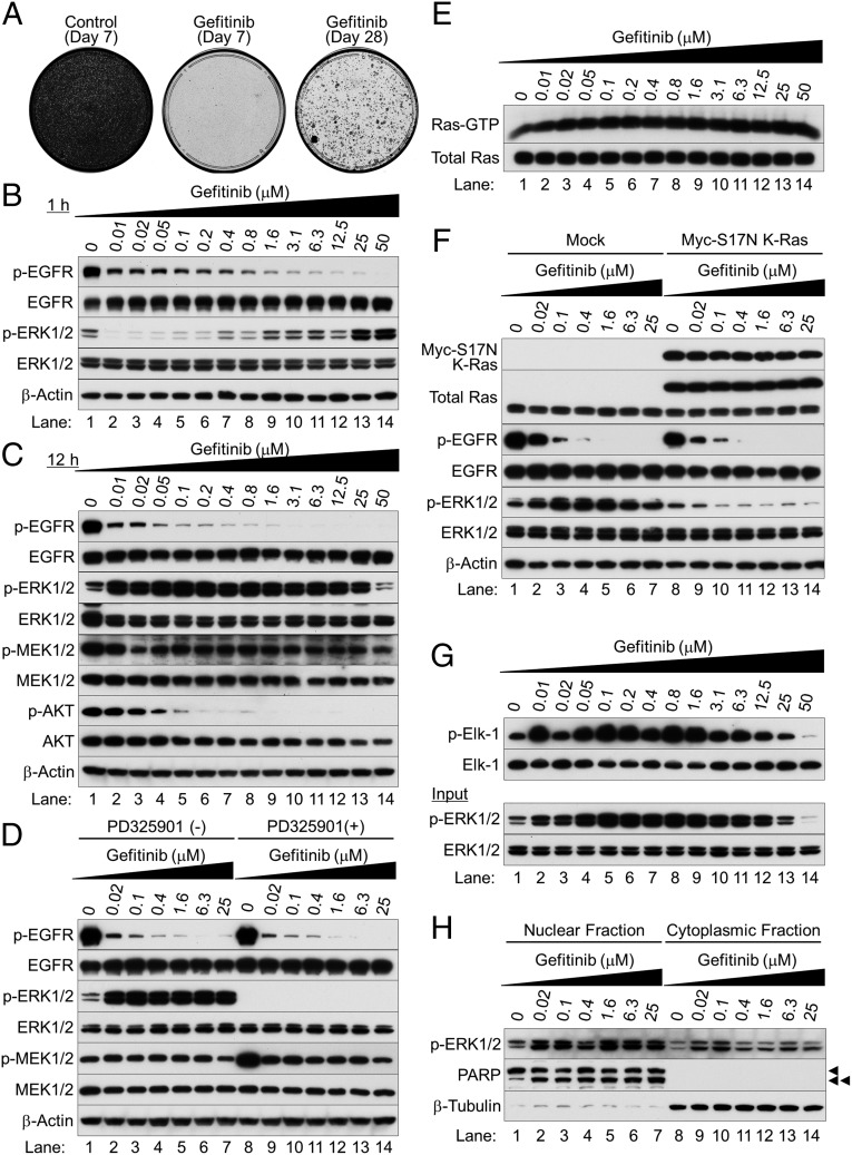
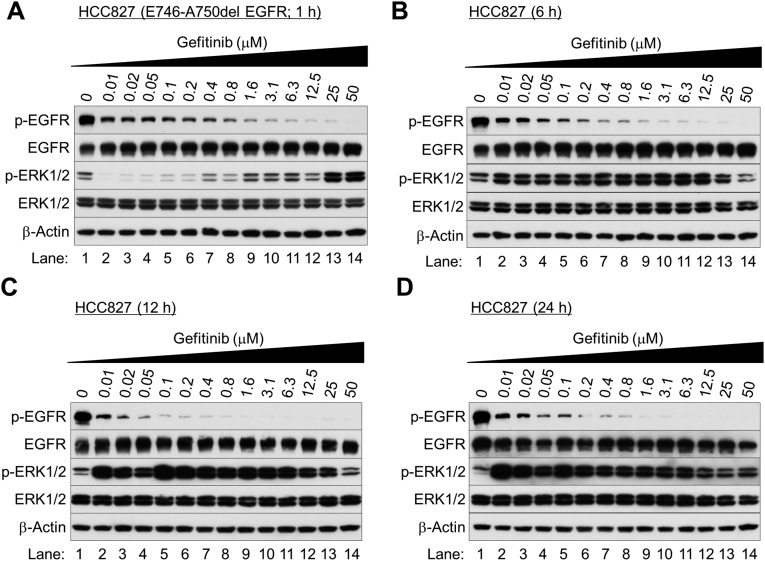
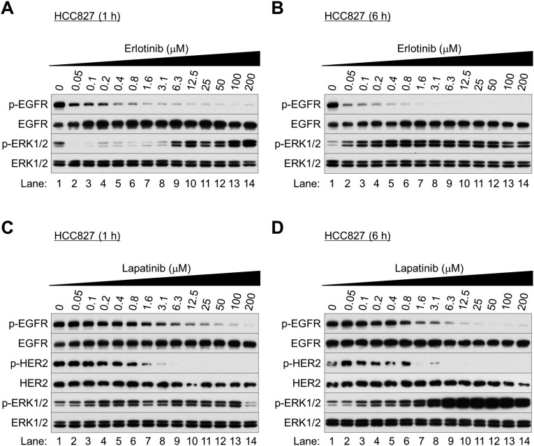
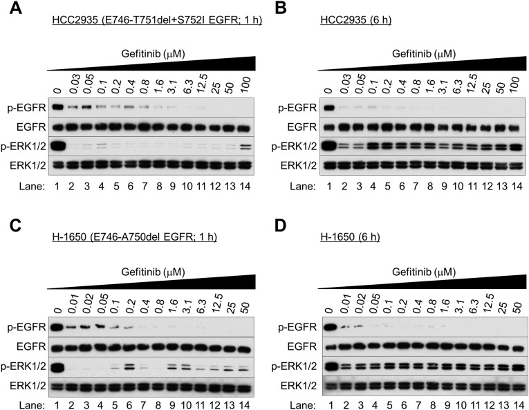
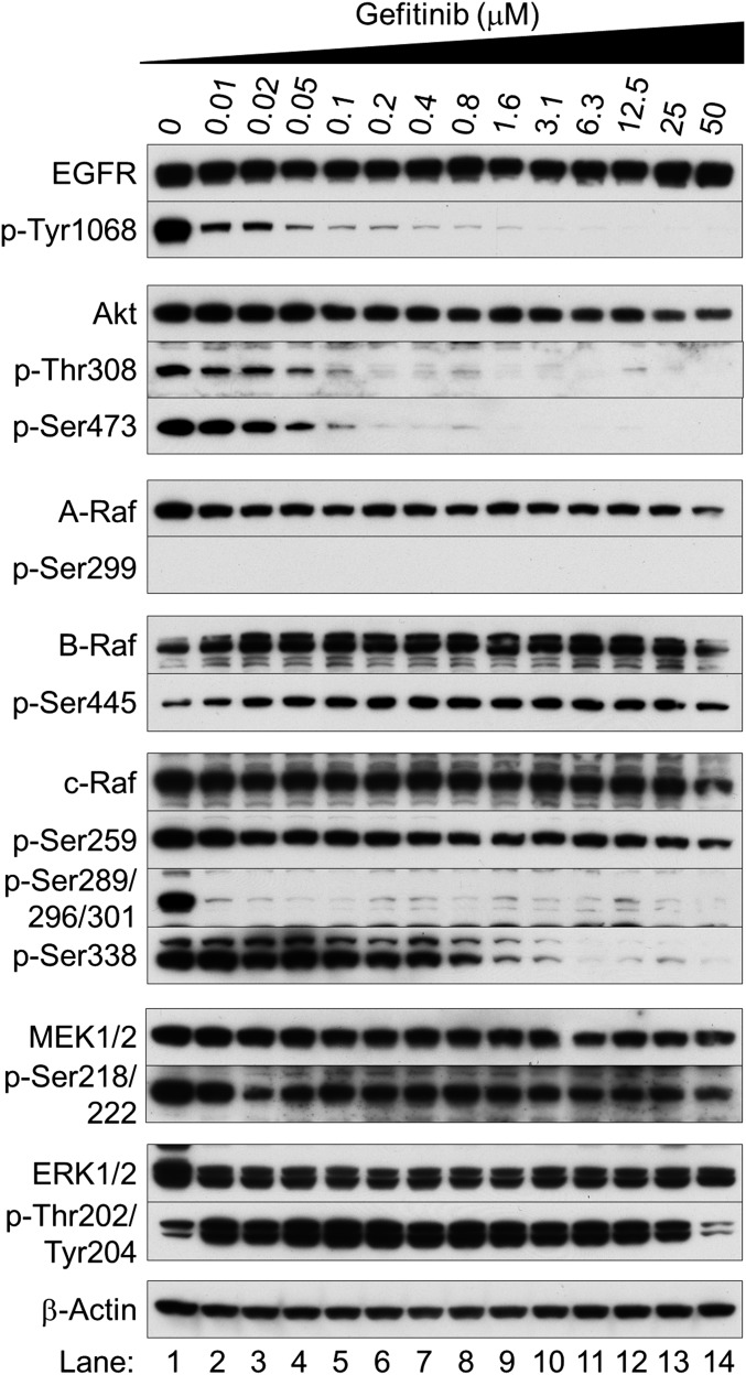
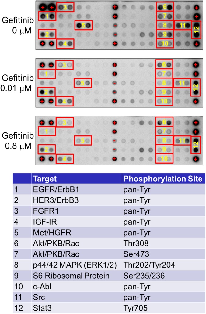
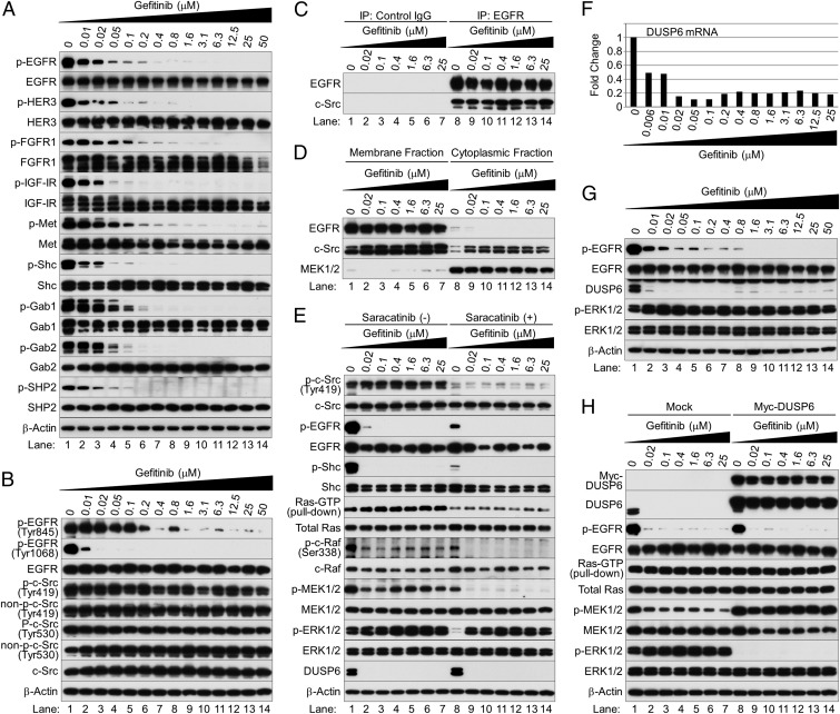
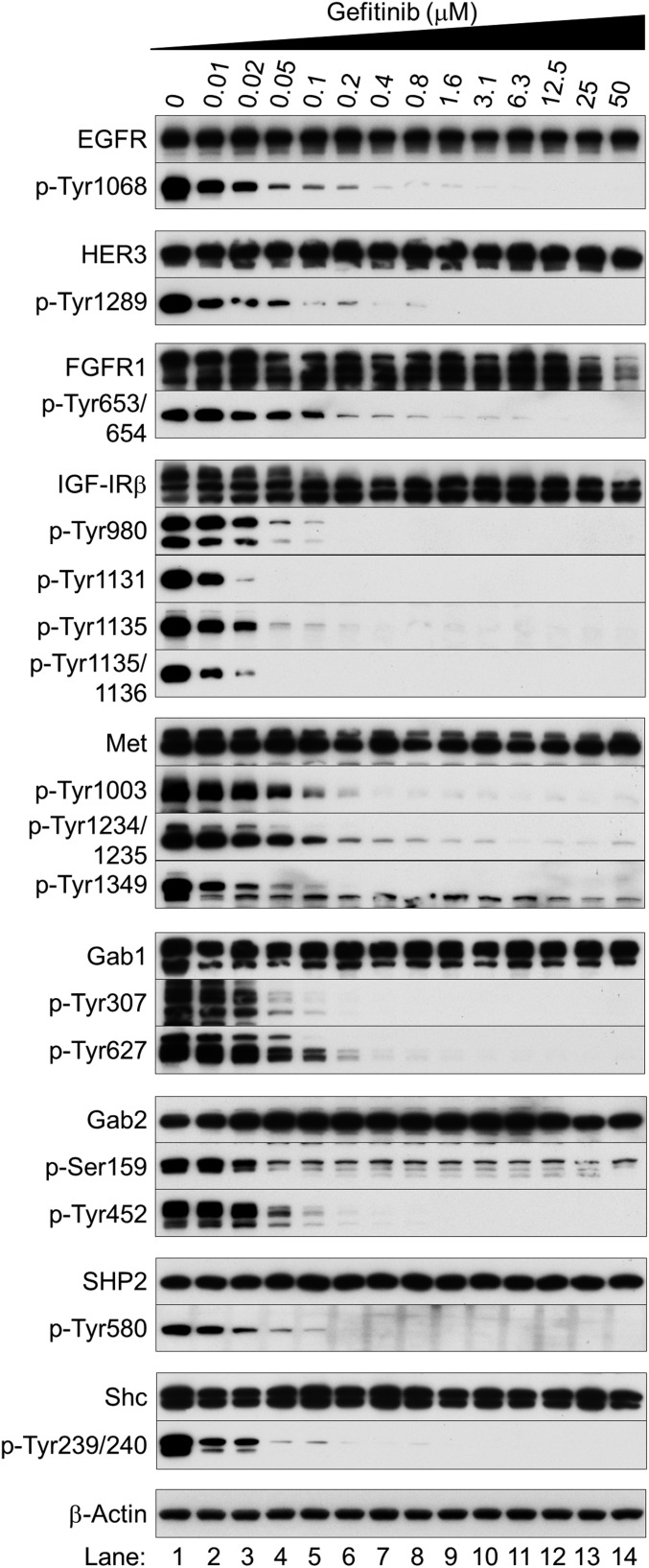
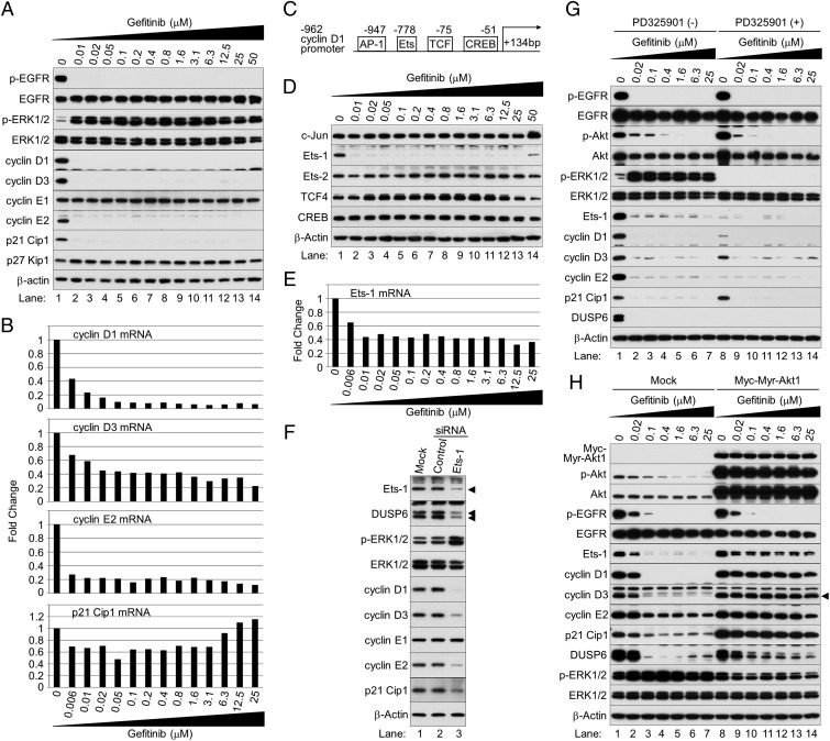
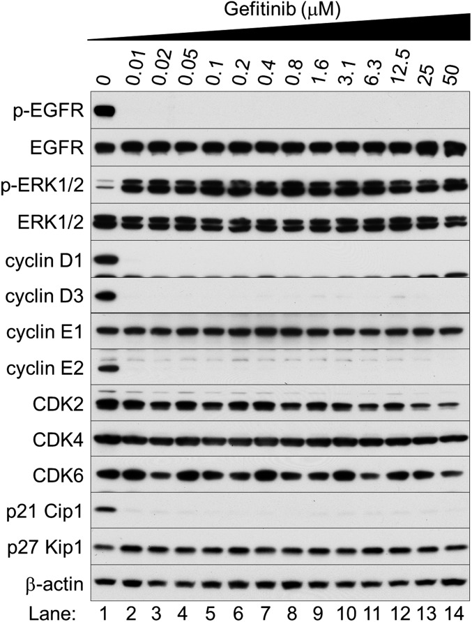
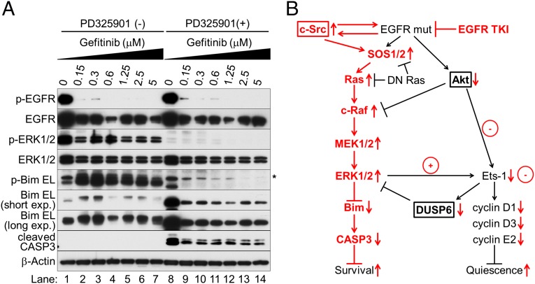
Nonsmall cell lung cancer (NSCLC) is the leading cause of cancer death worldwide. About 14% of NSCLCs harbor mutations in epidermal growth factor receptor (EGFR). Despite remarkable progress in treatment with tyrosine kinase inhibitors (TKIs), only 5% of patients achieve tumor reduction >90%. The limited primary responses are attributed partly to drug resistance inherent in the tumor cells before therapy begins. Recent reports showed that activation of receptor tyrosine kinases (RTKs) is an important determinant of this innate drug resistance. In contrast, we demonstrate that EGFR inhibition promotes innate drug resistance despite blockade of RTK activity in NSCLC cells. EGFR TKIs decrease both the mitogen-activated protein kinase (MAPK) and Akt protein kinase pathways for a short time, after which the Ras/MAPK pathway becomes reactivated. Akt inhibition selectively blocks the transcriptional activation of Ets-1, which inhibits its target gene, dual specificity phosphatase 6 (DUSP6), a negative regulator specific for ERK1/2. As a result, ERK1/2 is activated. Furthermore, elevated c-Src stimulates Ras GTP-loading and activates Raf and MEK kinases. These observations suggest that not only ERK1/2 but also Akt activity is essential to maintain Ets-1 in an active state. Therefore, despite high levels of ERK1/2, Ets-1 target genes including DUSP6 and cyclins D1, D3, and E2 remain suppressed by Akt inhibition. Reduction of DUSP6 in combination with elevated c-Src renews activation of the Ras/MAPK pathway, which enhances cell survival by accelerating Bim protein turnover. Thus, EGFR TKIs evoke innate drug resistance by preventing Akt activity and inactivating Ets-1 function in NSCLC cells.

Keywords: EGFR inhibition; ERK1/2 paradoxical activation; innate drug resistance; nonsmall cell lung cancer; tyrosine kinase inhibitors.


Conflict of interest statement

The authors declare no conflict of interest.

Figures

Fig. 1.
Fig. 1.
Gefitinib paradoxically activates ERK1/2 in EGFR-mutated NSCLC cells. (A) Colony-forming assay on HCC827 cells with (Middle and Right) or without (Left) 1 μM gefitinib for 7 d (Left and Middle) or 28 d (Right). Cells were stained with 0.5% crystal violet containing 20% ethanol. (B and C) Western blot analysis. Exponentially growing HCC827 cells were treated with various concentrations of gefitinib and harvested at 1 h (B) and 12 h (C) after treatment. (D) Western blot analysis. HCC827 cells were treated with gefitinib for 12 h; 10 μM PD325901 was added, and the cells were cultured for 1 h before harvesting. (E) Active Ras pull-down assay. Total Ras expression was separately determined by Western blot analysis. (F) Western blot analysis. Exponentially growing HCC827 cells were infected with lentivirus expressing Myc-S17N K-Ras or an empty vector; 48 h after infection, cells were cultured with or without gefitinib for 12 h. (G) Phospho-ERK1/2–associated GST-Elk1 in vitro kinase assay (Top). Western blots of total cell lysates (Bottom). (H) Subcellular fractionation analysis. Arrowheads indicate full-length PARP (single arrowhead) and cleaved PARP (double arrowhead).
Fig. S1.
Fig. S1.
Gefitinib paradoxically activates ERK1/2 in EGFR-mutated NSCLC cells after 6–24 h of incubation. Western blot analysis. Exponentially growing HCC827 cells were treated with various concentrations of gefitinib and collected after 1 h (A), 6 h (B), 12 h (C), and 24 h (D) of treatment.
Fig. S2.
Fig. S2.
EGFR TKIs paradoxically activate ERK1/2 in EGFR-mutated NSCLC cells. (AD) Western blot analysis. Exponentially growing HCC827 cells were treated with erlotinib (A and B) or lapatinib (C and D) at the indicated concentrations and collected after 1 h (A and C) and 6 h (B and D) of treatment.
Fig. S3.
Fig. S3.
Gefitinib paradoxically activates ERK1/2 in EGFR-mutated NSCLC cells. (AD) Western blot analysis. Exponentially growing HCC2935 cells (A and B) or H-1650 cells (C and D) were treated with gefitinib and collected after 1 h (A and C) and 6 h (B and D) of treatment.
Fig. S4.
Fig. S4.
Gefitinib paradoxically activates the MAPK pathway in EGFR-mutated NSCLC cells. Western blot analysis was performed with antibodies specific for the indicated phosphorylation sites and total antibodies. Exponentially growing HCC827 cells were treated with different concentrations of gefitinib and collected after 12 h of treatment.
Fig. S5.
Fig. S5.
Gefitinib paradoxically activates ERK1/2 in NSCLC cells bearing wild-type EGFR but not a drug-resistant mutant form after 6 h of incubation. (AF) Western blot analysis. Exponentially growing H-1838 cells (A and B; EGFR wild-type EGFR), H-2170 cells (C and D; EGFR wild-type), and H-1975 cells (E and F; T790M + L858R TKI-resistance mutant form of EGFR) cells were treated with various concentration of gefitinib and collected after 1 h (A, C, and E) and 6 h (B, D, and F) h of treatment.
Fig. S6.
Fig. S6.
Phosphorylation of EGFR, HER3, FGFR1, IGF1R, and Met was reduced in cells treated with gefitinib for 12 h. Phospho-RTK antibody arrays. The phosphorylation status of 28 major RTKs was simultaneously assessed.
Fig. 2.
Fig. 2.
c-Src activates the EGFR/MAPK pathway in NSCLC cells and cooperates with loss of DUSP6 to activate ERK1/2 after EGFR inhibition. (A and B) Western blot analysis. Cells were treated with gefitinib at the indicated concentrations for 12 h. (C) Immunoprecipitation and immunoblotting. Protein was precipitated with mouse antibodies specific to control IgG or EGFR, followed by immunoblotting. (D) Subcellular fractionation analysis. (E) Western blot analysis and active Ras pull-down assay. HCC827 cells were treated with gefitinib for 12 h and further cultured with saracatinib for 1 h before harvesting. (F and G) HCC827 cells were treated with various concentrations of gefitinib for 12 h. (F) Quantitative PCR. Fold changes in gene expression were calculated as the ratio of expression in each sample treated with different concentrations of gefitinib to the level without it. (G) Western blot analysis. (H) Western blot analysis and active Ras pull-down assay. Exponentially growing HCC827 cells were infected with a lentivirus expressing Myc-tagged DUSP6 or empty vector lentivirus (mock infection); 48 h after infection, cells were cultured in the presence or absence of gefitinib for 12 h and harvested.
Fig. S7.
Fig. S7.
RTKs are not responsible for Ras/MAPK activation induced by EGFR inhibitors. Western blot analysis was performed using antibodies to indicated specific phosphorylation sites or total expression. Exponentially growing HCC827 cells were treated with different concentrations of gefitinib and collected after 12 h of treatment.
Fig. 3.
Fig. 3.
Inhibition of Akt protein kinase after exposure to gefitinib is the primary cause of reduced expression of Ets-1, cyclins D1, D3, and E2, and DUSP6. (A) Western blot analysis of cyclin D1 and other G1 cell-cycle regulation molecules. (B) Quantitative PCR. Fold changes in gene expression were calculated as the ratio of expression in each sample treated with gefitinib to levels without it. (C) Schematic of −962 cyclin D1 promoter. (D) Western blot analysis of potential cyclin D1 regulation molecules. (E) Quantitative PCR of Ets-1. (F) Western blot. Ets-1 expression in HCC827 cells was depleted for 48 h with siRNA. Control: nontargeting siRNA. Mock: mock transfection. Arrowheads indicate Ets-1 and p42 and p44 DUSP6 proteins. (G) Western blot analysis. HCC827 cells were cultured in the presence or absence of different concentrations of gefitinib with or without PD325901 for 12 h. (H) Western blot analysis. Exponentially growing HCC827 cells were infected with a lentivirus expressing Myc-Myr-Akt1 or an empty vector; 48 h later, cells were cultured in the presence or absence of gefitinib for 12 h and harvested. The arrowhead shows cyclin D3-specific bands.
Fig. S8.
Fig. S8.
Gefitinib inhibits expression of cyclins D1, D3, and E2 but not cyclin E1, CDK 2, 4, and 6, and p27 Kip1. Western blot analysis of G1 cell-cycle regulation molecules.
Fig. S9.
Fig. S9.
HCC827 cells exclusively express TCF4 among TCF/LEF family transcription factors. Western blot analysis is shown. Cell lysates were separately prepared from the indicated exponentially growing cell lines. Arrowheads indicate 58- and 79-kDa TCF4 splice variants.
Fig. 4.
Fig. 4.
Sustained activation of ERK1/2 enhances cell survival by accelerating Bim protein turnover. (A) Western blot analysis. HCC827 cells were cultured in the presence or absence of different concentrations of gefitinib for 5 d; medium was then changed and cells were further cultured with or without 10 μM PD325901 for 24 h before harvesting. Asterisk stands for nonspecific bands. (B) Schematic of the molecular mechanism of EGFR inhibition and innate drug resistance in EGFR-mutated NSCLC cells.

Comment in

Similar articles

Cited by

References

    1. Pao W, Chmielecki J. Rational, biologically based treatment of EGFR-mutant non-small-cell lung cancer. Nat Rev Cancer. 2010;10(11):760–774. - PMC - PubMed
    1. Chong CR, Jänne PA. The quest to overcome resistance to EGFR-targeted therapies in cancer. Nat Med. 2013;19(11):1389–1400. - PMC - PubMed
    1. Rosell R, et al. Spanish Lung Cancer Group in collaboration with Groupe Français de Pneumo-Cancérologie and Associazione Italiana Oncologia Toracica Erlotinib versus standard chemotherapy as first-line treatment for European patients with advanced EGFR mutation-positive non-small-cell lung cancer (EURTAC): A multicentre, open-label, randomised phase 3 trial. Lancet Oncol. 2012;13(3):239–246. - PubMed
    1. Lippert TH, Ruoff HJ, Volm M. Current status of methods to assess cancer drug resistance. Int J Med Sci. 2011;8(3):245–253. - PMC - PubMed
    1. Wilson TR, et al. Widespread potential for growth-factor-driven resistance to anticancer kinase inhibitors. Nature. 2012;487(7408):505–509. - PMC - PubMed

Publication types

MeSH terms