. 2015 Jul 21;112(29):E3806-15.
doi: 10.1073/pnas.1505813112. Epub 2015 Jul 6.

Skip residues modulate the structural properties of the myosin rod and guide thick filament assembly

Affiliations

Skip residues modulate the structural properties of the myosin rod and guide thick filament assembly

Keenan C Taylor et al. Proc Natl Acad Sci U S A. .

Abstract

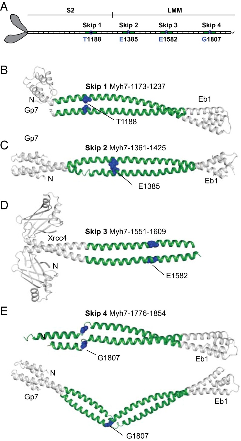
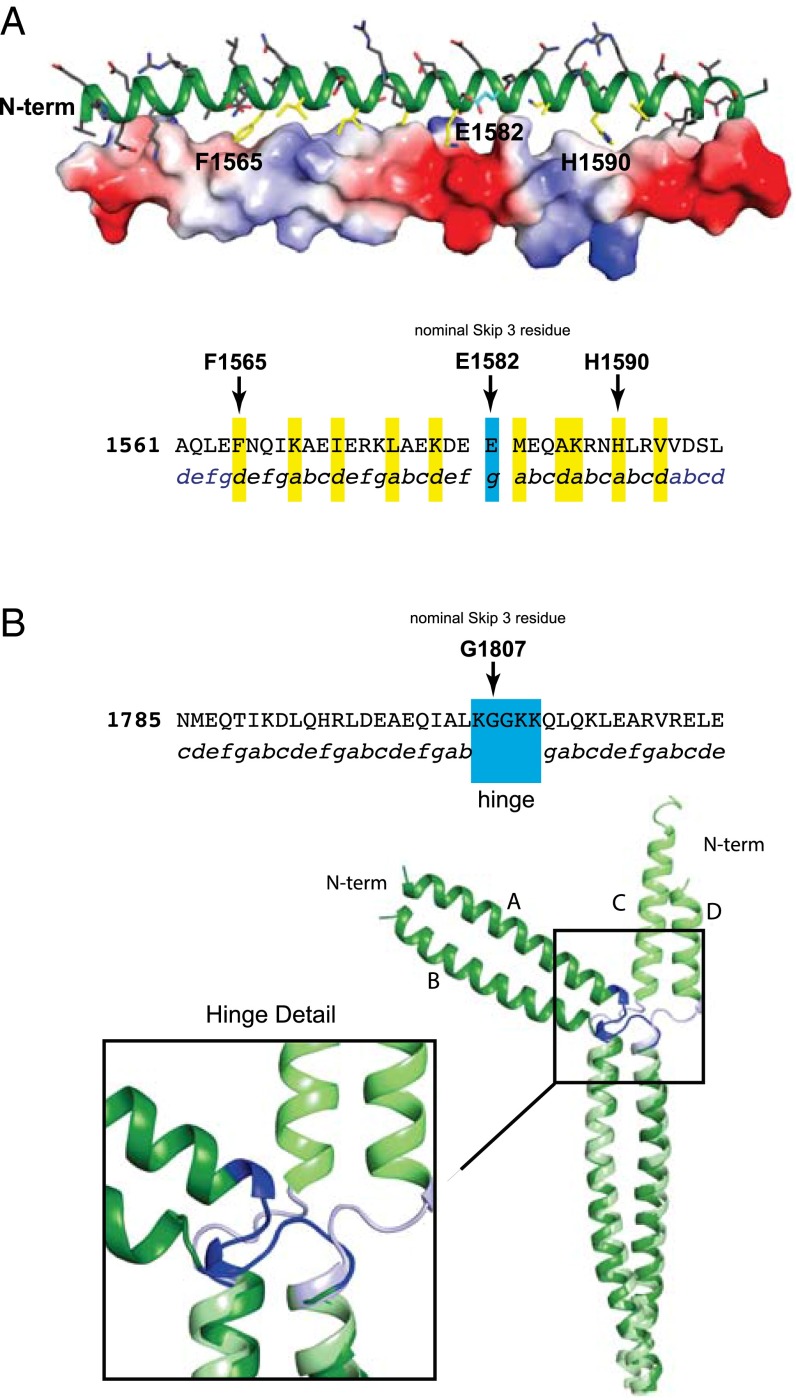
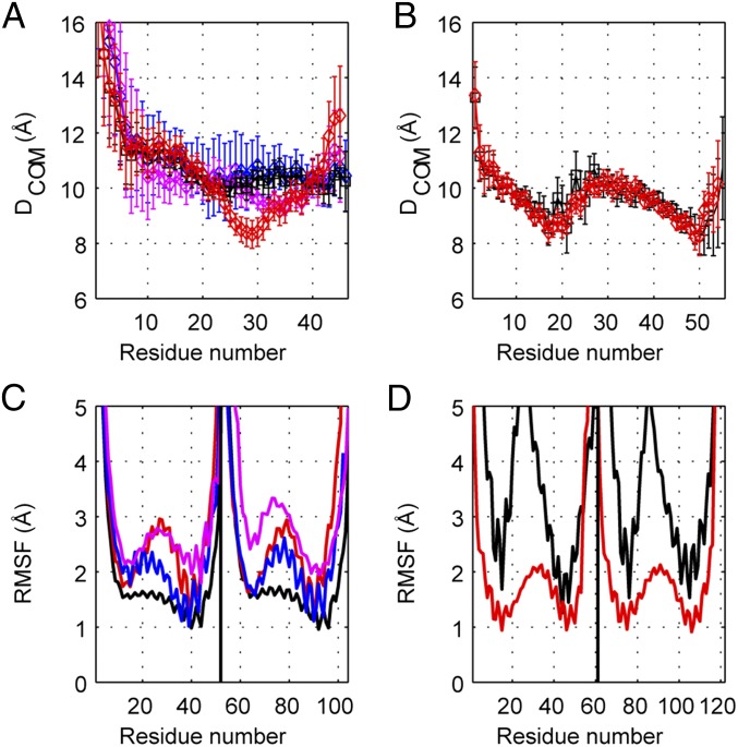
The rod of sarcomeric myosins directs thick filament assembly and is characterized by the insertion of four skip residues that introduce discontinuities in the coiled-coil heptad repeats. We report here that the regions surrounding the first three skip residues share high structural similarity despite their low sequence homology. Near each of these skip residues, the coiled-coil transitions to a nonclose-packed structure inducing local relaxation of the superhelical pitch. Moreover, molecular dynamics suggest that these distorted regions can assume different conformationally stable states. In contrast, the last skip residue region constitutes a true molecular hinge, providing C-terminal rod flexibility. Assembly of myosin with mutated skip residues in cardiomyocytes shows that the functional importance of each skip residue is associated with rod position and reveals the unique role of the molecular hinge in promoting myosin antiparallel packing. By defining the biophysical properties of the rod, the structures and molecular dynamic calculations presented here provide insight into thick filament formation, and highlight the structural differences occurring between the coiled-coils of myosin and the stereotypical tropomyosin. In addition to extending our knowledge into the conformational and biological properties of coiled-coil discontinuities, the molecular characterization of the four myosin skip residues also provides a guide to modeling the effects of rod mutations causing cardiac and skeletal myopathies.

Keywords: cardiac/skeletal myopathies; coiled-coils; molecular dynamics; myosin; protein structure.


Conflict of interest statement

The authors declare no conflict of interest.

Figures

Fig. 1.
Fig. 1.
Cartoon of their location in the myosin rod and structures of the four human β-cardiac myosin (MYH7) skip residues. (A) The relative location of the skip residues in the myosin rod which depicts the 38 dipolar charge repeats, each of which is formed by 28 amino acid residues (5). Each fusion protein consists of an N-terminal globular element, either Gp7 or Xrcc4 (white), before a section of MYH7 (green). A C-terminal fusion, Eb1 (white), is also present in all constructs except for Skip 3: Xrcc4-L1551-N1609. Each skip residue is colored in blue and depicted in sphere representation. The N terminus of each construct is indicated. (B) Gp7-K1173-I1238-Eb1 (Skip 1). (C) Gp7-L1361-I1425-Eb1 (Skip 2). (D) Xrcc4-L1551-N1609 (Skip 3). (E) Two crystallographically independent dimers within the asymmetric unit are shown for Gp7-A1777-T1854-Eb1 (Skip 4). Gp7 is disordered in the crystal lattice for the first of the two dimers shown in E.
Fig. 2.
Fig. 2.
Structural analysis of Skip 3 and Skip 4. (A, Upper) A cartoon representation of the coiled-coil surrounding E1582 (Skip 3) and with a surface electrostatic representation shown on the lower-helix. (Lower) The protein sequence surrounding Skip 3. Packing residues in the nonconical coiled-coil region of the upper-helix are colored in yellow on the upper-helix. The skip residue is colored in cyan. The protein sequence surrounding the nominal skip residue, E1582, with the observed coiled-coil position registry shown below. Residues that are in a standard packing arrangement are in blue and the atypical region is colored black. Residues packing along the distorted interface are highlighted in yellow and the skip residue is in cyan. (B) The sequence and structure of Skip 4. The protein sequence surrounding the nominal Skip 4 residue, G1807, is shown with the coiled-coil position registry below. Residues involved in the hinge region are indicated. A C-terminal structural alignment of residues Q1811–T1854 in chains A and B superimposed on C and D for the two independent molecules in the asymmetric unit for Skip 4 is also shown. This reveals the conformational variability in the Skip 4 hinge. The stabilization and folding domains were omitted from all structural figures.
Fig. 3.
Fig. 3.
Analysis of the molecular dynamics simulations for Skip 3 and Skip 4. DCOM and RMSF for the regions surrounding Skip 3 (A and C, respectively) and Skip 4 (B and D, respectively). The analyses of the WT simulations are shown in black, the skip residue deletion simulations (ΔS) are depicted in red. The simulation of the recoiled Skip 3 WT sequence (S3-R) in which the distribution of hydrophobic residues matches that expected for a canonical coiled coil, but still includes Skip 3 is shown in pink, whereas the recoiled Skip 3 deletion (ΔS3-R) is depicted in blue. DCOM is the distance between two α-helices calculated from the center of masses of Cα atoms for seven consecutive amino acids and is inversely correlated with the degree of coiling in the simulations of models. Cα-RMSF values are an indication of the degree of flexibility, as well as stability. Regions with higher RMSF values have larger degrees of flexibility. The measurements were averaged over the final 500 ns of each simulation to allow sufficient sampling for relaxation and ensure convergence of ensembles. Residue numbers correspond to L1551-D1602 and K1783-S1843 for Skip 3 and Skip 4, respectively. The recoiled WT sequence is: (LEHEEGKILRAQLEFNQIKAEIERLAAEVDEELEQAVRNHLRVVDSLQTSLD) where Skip 3 is underlined and the mutated residues are shown in bold.
Fig. 4.
Fig. 4.
Diversity of ensembles formed by the regions surround Skip 3 and 4 in the presence and absence of the skip residues. (A) Skip 3 WT, (B) Skip 3 deletion ΔS3, (C) Skip 4 WT, (D) Skip 4 deletion ΔS4, (E) recoiled Skip 3 WT sequence, and (F) recoiled Skip 3 deletion (ΔS3-R). In this figure, Cα-RMSD with respect to the representative member of the most populated cluster was plotted against simulation time. The color-coding represents the different clusters formed where blue depicts the dominating clusters. The Cα-alignments of representative members onto the crystal structure and model structure, for Skip 3 and Skip 4, respectively, are shown on the right where the representative and initial structures are shown in blue and gray, respectively. Skip residues in the WT crystal structures are depicted in green. The percentage of each cluster and the Cα-RMSD to initial structure is given under each structure.
Fig. 5.
Fig. 5.
Effects of skip residue deletions on myosin incorporation into sarcomeres. (A) Cardiomyocytes electroporated with WT and mutant GFP-tagged skip residue deletion constructs (ΔS) were imaged by confocal microscopy 96 h later. (Scale bar, 10 μm.) (B) Cardiomyocytes were cotransfected with mutant GFP- and WT mCherry-tagged constructs as indicated. Cells were imaged by confocal microscopy 96 h later. The two boxes in the WT and ΔS4 merge panels show the high magnification view of the sarcomeric I band and H zone (the latter corresponding to the bare zone) and the lack of colocalization between the mutant GFP- and the WT mCherry-tagged myosins. (Scale bar, 5 μm.) (C) Linescan analysis showing the relative intensity across the sarcomere of WT GFP and mCherry (Left) and ΔS4 GFP- and WT mCherry- tagged myosins (Right). Cells from three independent transfections were imaged and a total of 420 sarcomeres for each graph analyzed; data were obtained by averaging the two fluorescence signals. x axis: pixel distance (0.086 μm per pixel); y axis: fluorescence intensity. The location of the I-band and H-zone are reported. (D) Colocalization of ΔS4 GFP construct with the endogenous myosin, and time course incorporation into the sarcomeres. (Ab-F59): cardiomyocytes were transfected with WT or ΔS4 GFP-tagged myosin constructs; 96 h later cells were fixed and stained with F59 antimyosin primary antibody that recognizes only the myosin head domain, and the Alexa Fluor 568 secondary antibody with orange-red emission color. All panels are overlays of GFP and mCherry fluorescence signals. (36 h, 48 h): cardiomyocytes cotranfected with ΔS4 GFP- and WT mCherry-tagged myosin rod constructs were imaged by confocal microscopy 36 and 48 h later. (Scale bar, 5 μm.)
Fig. 6.
Fig. 6.
Functional activity of mutants carrying duplications of 28 amino acids encompassing the Skip 2 residue. (A, Upper) Topology of the duplications showing the number of amino acids separating the skip residues from each other or from the beginning/end of the myosin rod. (A, Lower) alignment of 28 amino acids surrounding the Skip 2 residue with the corresponding Skip 3 and 4 regions (replaced by the duplications). The observed coiled-coil position registry is shown above and below the sequences together with the conserved charge distribution for Skip 2 and 3. (B) Cardiomyocytes were transfected with GFP-tagged myosin constructs as indicated. (C) Cardiomyocytes were cotransfected with mutant GFP- and WT mCherry-tagged myosin constructs as indicated. The box in the S2-S4 Repl merge images shows the high magnification view of the of the sarcomeric I-band and H-zone. Cells in both B and C were imaged by confocal microscopy 96 h after transfection. (Scale bars, 5 μm.) (D) Linescan analysis showing the relative intensity across five sarcomeres of S2-S3 Repl GFP- and WT mCherry-tagged myosins (Left) and S2-S4 Repl GFP- and WT mCherry-tagged myosins (Right). x axis: pixel distance (0.086 μm per pixel); y axis: fluorescence intensity. The location of the I-band and H-zone are reported.

Similar articles

Cited by

References

    1. Craig R. Structure of A-segments from frog and rabbit skeletal muscle. J Mol Biol. 1977;109(1):69–81. - PubMed
    1. Gautel M. The sarcomeric cytoskeleton: Who picks up the strain? Curr Opin Cell Biol. 2011;23(1):39–46. - PubMed
    1. Craig R, Woodhead JL. Structure and function of myosin filaments. Curr Opin Struct Biol. 2006;16(2):204–212. - PubMed
    1. Al-Khayat HA, Kensler RW, Squire JM, Marston SB, Morris EP. Atomic model of the human cardiac muscle myosin filament. Proc Natl Acad Sci USA. 2013;110(1):318–323. - PMC - PubMed
    1. McLachlan AD, Karn J. Periodic charge distributions in the myosin rod amino acid sequence match cross-bridge spacings in muscle. Nature. 1982;299(5880):226–231. - PubMed

Publication types

MeSH terms

LinkOut - more resources