. 2015 Aug 15;195(4):1524-37.
doi: 10.4049/jimmunol.1500952. Epub 2015 Jul 6.

VprBP Is Required for Efficient Editing and Selection of Igκ+ B Cells, but Is Dispensable for Igλ+ and Marginal Zone B Cell Maturation and Selection

Affiliations

VprBP Is Required for Efficient Editing and Selection of Igκ+ B Cells, but Is Dispensable for Igλ+ and Marginal Zone B Cell Maturation and Selection

Victoria L Palmer et al. J Immunol. .

Abstract

B cell development past the pro-B cell stage in mice requires the Cul4-Roc1-DDB1 E3 ubiquitin ligase substrate recognition subunit VprBP. Enforced Bcl2 expression overcomes defects in distal VH-DJH and secondary Vκ-Jκ rearrangement associated with VprBP insufficiency in B cells and substantially rescues maturation of marginal zone and Igλ(+) B cells, but not Igκ(+) B cells. In this background, expression of a site-directed Igκ L chain transgene increases Igκ(+) B cell frequency, suggesting VprBP does not regulate L chain expression from a productively rearranged Igk allele. In site-directed anti-dsDNA H chain transgenic mice, loss of VprBP function in B cells impairs selection of Igκ editor L chains typically arising through secondary Igk rearrangement, but not selection of Igλ editor L chains. Both H and L chain site-directed transgenic mice show increased B cell anergy when VprBP is inactivated in B cells. Taken together, these data argue that VprBP is required for the efficient receptor editing and selection of Igκ(+) B cells, but is largely dispensable for Igλ(+) B cell development and selection, and that VprBP is necessary to rescue autoreactive B cells from anergy induction.


Figures

Figure 1
Figure 1
Enforced Bcl2 expression partially rescues lymphocyte cellularity in Vprbpfl/fl Cre+ mice. (A) Diagram of wild-type and conditional Vprbp alleles; mb1-Cre expression deletes exons 7–8 in mice homozygous for the conditional Vprbp alleles (Vprbpfl/fl) and causes B cell developmental arrest at the pro-B-to-pre-B cell transition. (B) Analysis of wild-type (WT) mice and Vprbpfl/fl mice lacking or containing the mb1-Cre (Cre+) and/or Eμ-2-22 Bcl2 (Bcl2+) transgenes. For each genotype, the total number of cells and lymphocytes was determined in the bone marrow (BM) and spleen (SPL). Data are represented as mean +−-SEM. Statistically significant differences are indicated for selected group comparisons.
Figure 2
Figure 2
Enforced Bcl2 expression partially rescues B cell development in Vprbpfl/fl Cre+ mice. (A) Lymphocytes from mice with the indicated genotype were analyzed by flow cytometry for the expression of different surface markers using gating strategies defined under each row. Developmental subsets identified by the staining pattern are shown at right with corresponding gates. The percentage of cells in each gate is shown for representative animals. A splenic pro-B cell-like population detected in Vprbpfl/fl Bcl2+ mice is identified with an asterisk. (B-C) The absolute number of cells in various B cell developmental subsets in the bone marrow (B) or spleen (C) defined by flow cytometry in panel A was determined for each of the indicated mouse genotypes. Data are represented as mean +/− SEM and are summarized in Supplemental Table 1). Statistically significant differences are indicated for selected group comparisons.
Figure 3
Figure 3
B cells developing in Vprbp fl/fl Cre+ Bcl2+ mice are mostly Igλ+. (A) Gated splenic CD19+ lymphocytes from mice with the genotypes shown in Fig. 2 were analyzed for Igκ and Igλ expression by flow cytometry. (B-C) The proportion (B) and absolute number (C) of Igκ+ and Igλ+ cells in the bone marrow and spleen for each of the indicated mouse genotypes was determined using flow cytometric data shown in panel (A). Proportions were calculated as the percentage of Igκ+ cells (black bar) or Igλ+ cells (gray bar) among total CD19+ light chain-expressing lymphocytes (i.e., Igκ+ + Igλ+). Data in panel C are represented as mean +/− SEM (see also Supplemental Table 1). Statistically significant differences are indicated for selected group comparisons.
Figure 4
Figure 4
Vprbp fl/fl Cre+ mice show inefficient distal V(D)J rearrangement in the Ig heavy and kappa light chain loci which is rescued by enforced Bcl2 expression. Genomic DNA prepared from bone marrow pre-B cells (B220+CD43IgM) or splenic Igκ+ B cells (10000, 2500, or 625 cell equivalents) sorted from mice with the indicated genotypes was subjected to PCR and Southern hybridization to detect the V(D)J rearrangements shown at right. Amplification of the non-rearranging CD14 locus was performed as a loading control.
Figure 5
Figure 5
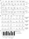
Loss of VprBP function does not alter intracellular Pax5 or Irf4 levels, but reduces cytoplasmic mu and kappa chain levels. (A-B) Gated bone marrow pro-B (B220+CD43+sIgM) and pre-B cells (B220+CD43sIgM) from mice with the genotypes shown in Fig. 2 were analyzed for the expression of (A) intracellular Pax5 and Irf4 or (B) cytoplasmic mu (cμ) and kappa (cκ) chains by flow cytometry. Representative staining by antigen-specific and isotype control antibodies (solid and dashed lines, respectively) is shown for animals of each genotype. In panel B, the additional shaded histogram represents isotype-specific staining of cells that were not permeabilized. Note that pro-B cells with a Pax5lo phenotype are CD19 whereas the Pax5hi cells are CD19+. The mean fluorescence intensity was calculated for n=5 animals/genotype and the data presented as the mean +/− SEM.
Figure 6
Figure 6
56R mice lacking functional VprBP express Ig heavy chain, but not Ig kappa chain, through early B cell developmental stages. (A-B) Bone marrow (BM) B cell developmental subsets identified by the staining pattern at right were analyzed for the expression of cytoplasmic mu heavy chain (cyto-μ; panel A) or kappa light chain (cyto-κ; panel B) and presented as in Fig. 5B. (C) The mean fluorescence intensity (MFI) of antigen-specific staining is summarized in Supplemental Fig. 1C.
Figure 7
Figure 7
Loss of VprBP function in 56R anti-dsDNA transgenic mice impairs receptor editing and selection of Ig kappa chains, but is dispensable for Ig lambda chain selection. (A) Splenocytes isolated from Vprbpfl/fl mice lacking or carrying the 56R, Cre, and/or Bcl2 transgenes in various combinations were analyzed for the expression of sIgMa (expressed from the 56R transgene) and sIgMb (expressed from the endogenous allele), Igκ and Igλ1/2 Igλx and Igλ1/2, or Igκ and Igλx on CD19+ lymphocytes using flow cytometry. The absolute numbers of CD19+ B cells expressing these markers are shown for each genotype in Supplemental Fig. 1A and Supplemental Table 1. (B) The proportion of CD19+ B cells expressing the markers examined in panel (A) were determined and presented as in Fig. 3B. Statistically significant differences are indicated for selected group comparisons.
Figure 8
Figure 8
Loss of VprBP function in 56R mice increases anergic B cell populations and impairs B cell selection into the marginal zone compartment. (A) Flow cytometry was used to identify various splenic transitional and mature B cell subsets as in Fig. 2A; the absolute numbers of cells in each subset are summarized in Supplemental Fig. 1B and Supplemental Table 1. A splenic pro-B cell-like population detected in Vprbpfl/fl Bcl2+ mice (identified with an asterisk) is notably absent in Vprbpfl/fl 56R+Bcl2+ mice. (B) Cytoplasmic mu heavy chain (cyto-μ) or kappa light chain (cyto-κ) was analyzed by flow cytometry in the populations identified in panel A and presented as in Fig. 5B. Examples of cyto-μ and cyto-κ staining profiles are shown for non-conventional B cell populations identified by an asterisk in panel A (transitional sIgMCD23 and mature CD21CD23+, respectively). The mean fluorescence intensity (MFI) of antigen-specific staining is summarized in Supplemental Fig. 1C. (C) The proportion of marginal zone (MZ) and follicular mature (FM) B cells in the spleen for each of the indicated mouse genotypes was determined using flow cytometric data shown in panel (A). Proportions were calculated as the percentage of FM cells (black bar) or MZ cells (gray bar) among total gated FM and MZ cells (i.e., FM + MZ). Statistically significant differences are indicated for selected groups expressing the 56R transgene.
Figure 9
Figure 9
Enforced 3-83κ light chain expression promotes development of Igκ+ B cells in Vprbpfl/fl Cre+ Bcl2+ mice. (A) Splenocytes isolated from Vprbp fl/fl mice lacking or carrying the 3-83κ, Cre, and/or Bcl2 transgenes in various combinations were analyzed for the expression of Igκ and Igλ on CD19+ lymphocytes using flow cytometry. (B) The proportion and absolute number of CD19+ B cells expressing Igκ or Igλ was determined and presented as in Fig. 3B-C. Statistically significant differences are indicated for selected groups expressing the 3-83κ transgene. (C) Genomic DNA prepared from total bone marrow (10000, 2500, or 625 cell equivalents) with the indicated genotypes was subjected to PCR and Southern hybridization to detect IRS-RS and Vλ1-to-Jλ1 rearrangements. Amplification of the non-rearranging CD14 locus was performed as a loading control.
Figure 10
Figure 10
Enforced 3-83κ light chain expression promotes development of Igκ+ B cells in Vprbpfl/fl Cre+ Bcl2+ mice. (A-C) Flow cytometry was used to identify various splenic transitional (panels A and B) and mature (panels A and C) B cell subsets as in Fig. 2A; the absolute numbers of cells in each subset are summarized in Supplemental Fig. 2C and Supplemental Table 2. A splenic pro-B cell-like population detected in Vprbpfl/fl Bcl2+ mice (identified with an asterisk) is notably present in Vprbpfl/fl 3-83κ+Bcl2+ mice. Conventional and non-conventional splenic transitional (panel B) and mature (panel C) B cell subsets were analyzed for cytoplasmic mu heavy chain (cyto-μ) or kappa light chain (cyto-κ) using flow cytometry as in Fig. 5B.

Similar articles

Cited by

References

    1. Schatz DG, Swanson PC. V(D)J Recombination: Mechanisms of Initiation. Annual Review Genetics. 2011;45:167–202. - PubMed
    1. Nakagawa T, Mondal K, Swanson PC. VprBP (DCAF1): a promiscuous substrate recognition subunit that incorporates into both RING-family CRL4 and HECT-family EDD/UBR5 E3 ubiquitin ligases. BMC Mol Biol. 2013;14:22. - PMC - PubMed
    1. Kassmeier MD, Mondal K, Palmer VL, Raval P, Kumar S, Perry GA, Anderson DK, Ciborowski P, Jackson S, Xiong Y, Swanson PC. VprBP binds full-length RAG1 and is required for B-cell development and V(D)J recombination fidelity. Embo J. 2012;31:945–958. - PMC - PubMed
    1. Brunner C, Marinkovic D, Klein J, Samardzic T, Nitschke L, Wirth T. B cell-specific transgenic expression of Bcl2 rescues early B lymphopoiesis but not B cell responses in BOB.1/OBF.1-deficient mice. J Exp Med. 2003;197:1205–1211. - PMC - PubMed
    1. Sitte S, Glasner J, Jellusova J, Weisel F, Panattoni M, Pardi R, Gessner A. JAB1 is essential for B cell development and germinal center formation and inversely regulates Fas ligand and Bcl6 expression. J Immunol. 2012;188:2677–2686. - PubMed

Publication types

MeSH terms

Substances

LinkOut - more resources