. 2015 Aug 15;195(4):1427-35.
doi: 10.4049/jimmunol.1500496. Epub 2015 Jul 6.

The B Cell-Stimulatory Cytokines BLyS and APRIL Are Elevated in Human Periodontitis and Are Required for B Cell-Dependent Bone Loss in Experimental Murine Periodontitis

Affiliations

The B Cell-Stimulatory Cytokines BLyS and APRIL Are Elevated in Human Periodontitis and Are Required for B Cell-Dependent Bone Loss in Experimental Murine Periodontitis

Toshiharu Abe et al. J Immunol. .

Abstract

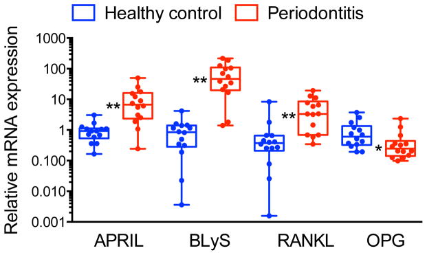
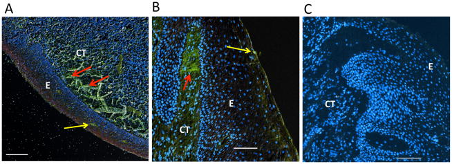
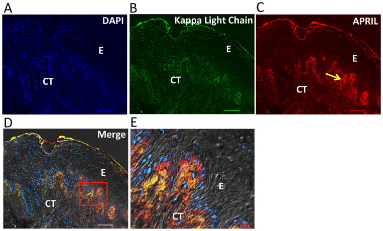
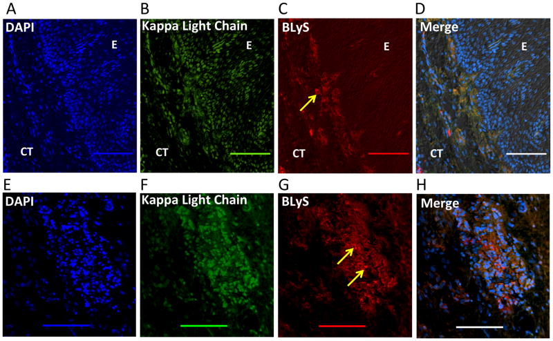
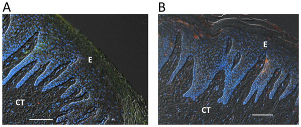
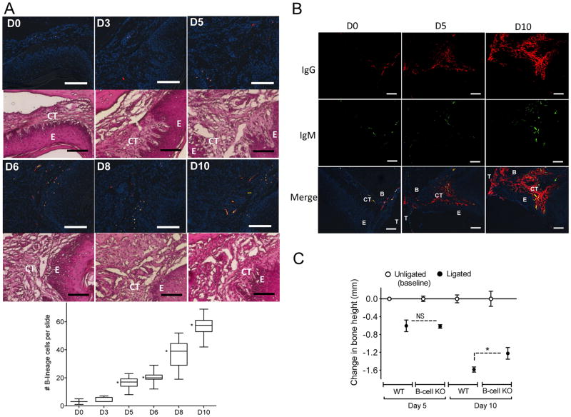
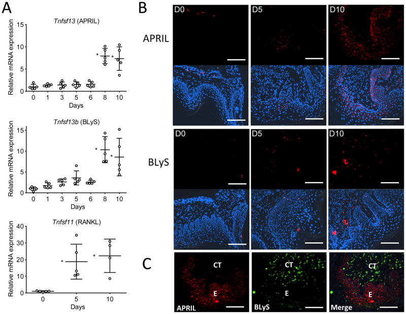
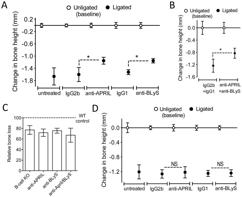
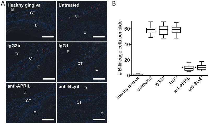
B-lineage cells (B lymphocytes and plasma cells) predominate in the inflammatory infiltrate of human chronic periodontitis. However, their role in disease pathogenesis and the factors responsible for their persistence in chronic lesions are poorly understood. In this regard, two cytokines of the TNF ligand superfamily, a proliferation-inducing ligand (APRIL) and B-lymphocyte stimulator (BLyS), are important for the survival, proliferation, and maturation of B cells. Thus, we hypothesized that APRIL and/or BLyS are upregulated in periodontitis and contribute to induction of periodontal bone loss. This hypothesis was addressed in both human and mouse experimental systems. We show that, relative to healthy controls, the expression of APRIL and BLyS mRNA and protein was upregulated in natural and experimental periodontitis in humans and mice, respectively. The elevated expression of these cytokines correlated with increased numbers of B cells/plasma cells in both species. Moreover, APRIL and BLyS partially colocalized with κ L chain-expressing B-lineage cells at the epithelial-connective tissue interface. Ligature-induced periodontitis resulted in significantly less bone loss in B cell-deficient mice compared with wild-type controls. Ab-mediated neutralization of APRIL or BLyS diminished the number of B cells in the gingival tissue and inhibited bone loss in wild-type, but not in B cell-deficient, mice. In conclusion, B cells and specific cytokines involved in their growth and differentiation contribute to periodontal bone loss. Moreover, APRIL and BLyS have been identified as potential therapeutic targets in periodontitis.


Figures

Figure 1
Figure 1. Upregulation of APRIL and BLyS mRNA expression in gingival biopsy specimens from periodontitis patients relative to healthy controls
Gingival mRNA expression of APRIL, BLyS, RANKL, and OPG (the latter two molecules were included for comparative reasons) in healthy and diseased sites was determined by qPCR. Results were normalized against GAPDH mRNA and presented as fold change in the transcript levels in diseased sites relative to those of healthy sites, which were assigned an average value of 1. Dots represent individual values (n = 14 subjects per group) and each box in the box and whisker plots extends from the 25th to the 75th percentile. *P < 0.05 and **P < 0.01 compared with healthy control.
Figure 2
Figure 2. Ig kappa light chain staining of human mucosal tissues
(A) Immunohistochemical analysis of human tonsil shows diffuse staining of kappa light chain (green) within the connective tissue indicating the presence of B cells/plasma cells (red arrows). (B) Immunohistochemical analysis of human gingival tissue from a patient with chronic periodontitis shows prominent staining of kappa light chain (green) within the connective tissue adjacent to the basement membrane (red arrow). In both the tonsil (A) and the gingiva (B), occasional kappa-light-chain–positive cells infiltrate the superficial epithelial layer (yellow arrows). (C) Control staining of human gingival tissue from a patient with chronic periodontitis using FITC-conjugated non-immune IgG. Nuclear staining (blue) by DAPI. E, epithelium; CT, connective tissue. Original magnification 100X. Scale bars, 100 μm.
Figure 3
Figure 3. APRIL expression in human periodontitis
Immunohistochemical analysis of diseased gingival tissue stained as follows: (A) DAPI to indicate cell nuclei. (B) Anti-kappa light chain mAb indicating the presence of Ig-expressing/secreting cells (B cells/plasma cells) predominantly within the gingival connective tissue directly adjacent to the basement membrane. (C) Anti-APRIL mAb showing immunoreactivity (yellow arrow) within the gingival epithelium and underlying connective tissue adjacent to the basement membrane. (D) Fluorescence and differential interference contrast merged image showing positive co-localization (orange/yellow) of APRIL and kappa light chain expression within the gingival connective tissue. (E) The demarcated area in D showing co-localized APRIL and kappa light chain surrounding DAPI-stained cells likely representing B cells/plasma cells. E, epithelium; CT, connective tissue. Original magnification 100X. Scale bars, 100 μm.
Figure 4
Figure 4. BLyS expression in human periodontitis
(A–D) Immunohistochemical analysis of diseased gingival tissue stained with as follows: (A) DAPI to indicate cell nuclei. (B) Anti-Ig kappa light chain mAb indicating the presence of Ig-expressing/secreting cells (B cells/plasma cells) predominantly within the gingival connective tissue. (C) Anti-BLyS mAb showing immunoreactivity (yellow arrow) within the gingival connective tissue and to a lesser extent within the epithelium. (D) Fluorescence and differential interference contrast merged image showing positive co-localization (orange) of BLyS and Ig kappa light chain expression within the gingival connective tissue. (E–H) Similar analysis as above using granulomatous tissue taken from the deepest aspect of an osseous defect. (E) DAPI staining to indicate cell nuclei. (F) Staining with anti-Ig kappa light chain mAb indicating the presence of Ig-expressing/secreting cells (B cells/plasma cells). (G) Staining with anti-BLyS mAb showing intense immunoreactivity (yellow arrows). (H) Fluorescence and differential interference contrast merged image showing intense positive co-localization (orange) of BLyS and Ig kappa light chain at heavily populated area of DAPI-stained cells likely representing B cells/plasma cells. E, epithelium; CT, connective tissue. Original magnification 200X. Scale bars, 100 μm.
Figure 5
Figure 5. APRIL and BLyS expression in healthy human gingival tissue
Immunohistochemical analysis of healthy gingiva stained for (A) APRIL or (B) BLyS (both red). In both A and B, the tissue was stained with DAPI to indicate cell nuclei (blue) and anti-Ig kappa light chain (green). The fluorescent images (merged with differential interference contrast) show little if any positive staining for APRIL, BLyS, or Ig kappa light chain (compare with diseased tissue in Figures 3 and 4). E, epithelium; CT, connective tissue. Original magnification 100X. Scale bars, 100 μm.
Figure 6
Figure 6. Presence and impact of B cells in murine periodontitis
(A and B) Tissue sections from ligature-induced periodontitis at the indicated timepoints were stained for CD19 (B cells; red) and CD138 (plasma cells; green) (A) or for mouse Igs as indicated (B). All immunofluorescent sections in A and the bottom row in B were stained with DAPI to indicate cell nuclei. E, epithelium; CT, connective tissue; T, tooth; B, bone. Scale bars, 100 μm. Original magnification 200X (A) and 100X (B). In A (left panel), H&E staining of the same tissue sections used for immunofluorescence analysis confirmed that the B-lineage cells are predominantly found in the connective tissue in proximity to the interface with the epithelium. In A (right panel), B-lineage cells were enumerated from 50 random sections (10 from each of five mice/time point) and data are presented as box-and-whisker plots. The median is marked inside each box, and the ends of each box correspond to the interquartile range from the 25th to the 75th percentile. *P < 0.01 as compared to D0. *P < 0.01 as compared to D0. (C) Periodontal bone loss was induced in groups of WT or B-cell KO mice for 5 or 10 d by ligating a maxillary second molar and leaving the contralateral tooth unligated (baseline control). Data are means ± SD (n = 5 mice per group). *P < 0.01 between indicated groups. NS, not significant.
Figure 7
Figure 7. Expression of APRIL and BLyS in the murine gingival tissue
(A) Timecourse of APRIL (upper), BLyS (middle), and RANKL (lower) mRNA expression in the gingival tissue after ligature-induced periodontitis for 10 d in mice. Results were normalized to GAPDH mRNA and presented relative to those at day 0, set as 1. Scatter dots and mean ± SD were plotted for data from each group of mice (n = 5 mice per timepoint). *P < 0.01 compared to day 0. (B) Gingival tissue sections from ligature-induced periodontitis at days 0, 5, and 10 (D0, D5, and D10) were stained for APRIL (upper panels) or BLyS (lower panels). The APRIL and BLyS fluorescent images were merged with DAPI staining (blue) to reveal nuclei. (C) Staining for APRIL and BLyS in a gingival tissue section at day 10 post-ligation and merged image. E, epithelium; CT, connective tissue. Original magnification 200X. Scale bars, 100 μm.
Figure 8
Figure 8. Ab-mediated neutralization of APRIL or BLyS inhibits ligature-induced periodontal bone loss in a B cell-dependent manner
(A) Periodontal bone loss in WT mice locally microinjected (or not) with 5 μg anti-APRIL mAb or anti-BLyS mAb (or equal amounts of their isotype controls), 4 d after placing the ligatures and every day thereafter until the day before sacrifice (day 10). (B) Similar bone loss experiment in which mice were microinjected with a combination of anti-APRIL mAb and anti-BLyS mAb or a combination of their isotype controls (each mAb or control was used at 5 μg). (C) The bone loss for each treatment in A and B was calculated relative to their unligated controls (set as 100%). Also included for comparison is the bone loss of B-cell KO relative to WT controls from the experiment in figure 6C. (D) Periodontal bone loss in B-cell KO mice locally microinjected (or not) with 5 μg anti-APRIL mAb or anti-BLyS mAb (or equal amounts of their isotype controls), 4 d after placing the ligatures and every day thereafter until the day before sacrifice (day 10). Data are means ± SD (n = 5 mice per group). Data are means ± SD (n = 5 mice per group). *P < 0.01 between indicated groups. NS, not significant.
Figure 9
Figure 9. Ab-mediated neutralization of APRIL or BLyS diminishes the numbers of B cells in the gingival tissue of mice subjected to ligature-induced periodontitis
(A) Mice undergoing ligature-induced periodontitis were locally microinjected or not (“untreated”) with 5 μg anti-APRIL mAb or anti-BLyS mAb (or equal amounts of their isotype controls, IgG2b and IgG1, respectively), 4 d after placing the ligatures and every day thereafter until the day before sacrifice (day 10). Gingival tissue sections from these groups of mice as well as from mice not subjected to ligature-induced periodontitis (“healthy gingiva”) were stained for CD19 (B cells; red) and DAPI (blue) to reveal nuclei. E, epithelium; CT, connective tissue; B, bone. Scale bars, 100 μm. Original magnification 200X. (B) B cells were enumerated from 30 random sections (10 from each of three mice/group) and data are presented as box-and-whisker plots. The median is marked inside each box, and the ends of each box correspond to the interquartile range from the 25th to the 75th percentile. *P < 0.01 as compared to untreated and corresponding isotype control.

Comment in

References

    1. Hajishengallis G. Periodontitis: from microbial immune subversion to systemic inflammation. Nat Rev Immunol. 2015;15:30–44. - PMC - PubMed
    1. Lamont RJ, Hajishengallis G. Polymicrobial synergy and dysbiosis in inflammatory disease. Trends Mol Med. 2015;21:172–183. - PMC - PubMed
    1. Hajishengallis G. Immunomicrobial pathogenesis of periodontitis: keystones, pathobionts, and host response. Trends Immunol. 2014;35:3–11. - PMC - PubMed
    1. Garlet GP. Destructive and protective roles of cytokines in periodontitis: A reappraisal from host defense and tissue destruction viewpoints. J Dent Res. 2010;89:1349–1363. - PubMed
    1. Gemmell E, Yamazaki K, Seymour GJ. The role of T cells in periodontal disease: homeostasis and autoimmunity. Periodontol 2000. 2007;43:14–40. - PubMed

Publication types

MeSH terms

Substances

LinkOut - more resources