. 2015 Sep;83(9):3648-56.
doi: 10.1128/IAI.00545-15. Epub 2015 Jul 6.

Heparan Sulfate Modulates Neutrophil and Endothelial Function in Antibacterial Innate Immunity

Affiliations

Heparan Sulfate Modulates Neutrophil and Endothelial Function in Antibacterial Innate Immunity

Ding Xu et al. Infect Immun. 2015 Sep.

Abstract

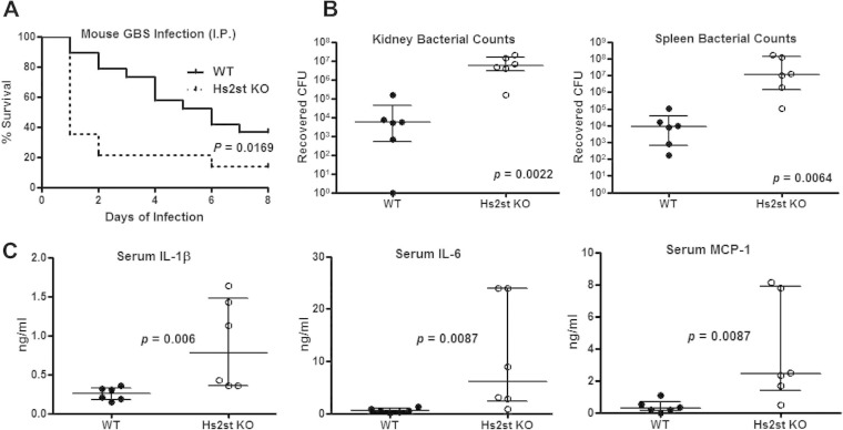
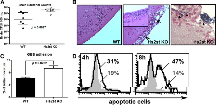
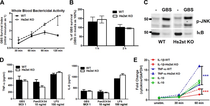
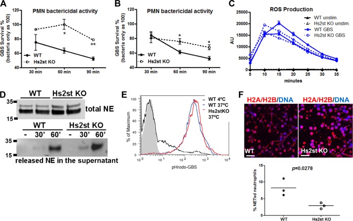
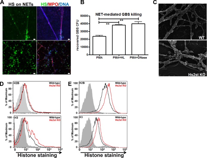
Recently, we showed that endothelial heparan sulfate facilitates entry of a bacterial pathogen into the central nervous system. Here, we show that normal bactericidal activity of neutrophils is influenced by the sulfation pattern of heparan sulfate. Inactivation of heparan sulfate uronyl 2-O-sulfotransferase (Hs2st) in neutrophils substantially reduced their bactericidal activity, and Hs2st deficiency rendered mice more susceptible to systemic infection with the pathogenic bacterium group B Streptococcus. Specifically, altered sulfation of heparan sulfate in mutant neutrophils affected formation of neutrophil extracellular traps while not influencing phagocytosis, production of reactive oxygen species, or secretion of granular proteases. Heparan sulfate proteoglycan(s) is present in neutrophil extracellular traps, modulates histone affinity, and modulates their microbial activity. Hs2st-deficient brain endothelial cells show enhanced binding to group B Streptococcus and are more susceptible to apoptosis, likely contributing to the observed increase in dissemination of group B Streptococcus into the brain of Hs2st-deficient mice following intravenous challenge. Taken together, our data provide strong evidence that heparan sulfate from both neutrophils and the endothelium plays important roles in modulating innate immunity.


Figures

FIG 1
FIG 1
Exacerbated mortality and bacterial dissemination in Hs2St-deficient mice. (A) Increased mortality of Hs2St-deficient mice after GBS challenge. Survival of mice was monitored for 8 days after intraperitoneal (i.p.) infection with 1.5 × 10⁸ CFU GBS. Data were pooled from two independent experiments (n = 19 for WT and n = 14 for Hs2St-deficient mice). Differences between groups were calculated by a log rank test. Mice were infected i.p. with 5 × 10⁷ CFU of GBS and then euthanized at 18 h postinfection for collection of organs for enumeration of bacterial CFU (B) and inflammatory cytokine ELISA (C). Data are medians with interquartile ranges from one of the two independent experiments. Each circle denotes one mouse (n = 6/group). Differences between groups were calculated by Mann-Whitney test (B and C).
FIG 2
FIG 2
Increased GBS association and apoptosis of the Hs2st-deficient endothelial cells contributed to the GBS brain dissemination. Mice were intravenously challenged with 1.4 × 10⁸ CFU of GBS, and brains were collected 18 h after GBS infection to measure bacterial loads (A) or fixed in formalin for H&E staining (B, left and middle; note the damage to the meninges) and Gram staining (B, right). Data are medians with interquartile ranges from one of the two independent experiments, and each symbol denotes 1 mouse (n = 6 for each group). (C) Increased GBS association with Hs2st-deficient mouse brain microvascular endothelial cells (BMECs). Primary isolated BMECs were infected with GBS (MOI = 10) for 30 min, followed by extensive washing to remove unbound GBS. Cell-associated GBS were enumerated by serial plating. Differences between the two groups were calculated by unpaired t test. (D) BMECs were infected with GBS at an MOI of 10 for the indicated times, and apoptotic cells were detected by TUNEL assay. Representative data are from one of the 2 or 3 independent experiments (C and D).
FIG 3
FIG 3
Hs2st-deficient macrophages are competent to respond to bacterial challenge. (A) Increased replication of GBS in whole blood from Hs2St-deficient mice. GBS (104 CFU) were incubated with blood collected from WT or Hs2St-deficient mice, and surviving bacteria were enumerated at the indicated time points by serial plating. Data are representative of two independent experiments. (B) Similar bactericidal activity between WT and Hs2st-deficient macrophages. Mouse bone marrow-derived macrophages (MBDMs) were infected with GBS at an MOI of 0.2, and surviving GBS were enumerated at the indicated time point by serial plating. (C) MBDMs from WT and Hs2st-deficient mice were challenged with GBS for 30 min, and cell lysates were collected to analyze IκB degradation and JNK phosphorylation by Western blotting. (D) Macrophage cytokine secretion upon bacterial challenge. MBDMs were challenged with GBS, Pam2CKS4, and LPS for 24 h, and the culture supernatant from the stimulated macrophages was harvested for cytokine ELISA. (E) mRNA was collected from WT and Hs2st-deficient macrophages after GBS stimulation to analyze cytokine transcript production by quantitative RT-PCR. Data are means ± standard errors of the means (SEM) from one of 2 or 3 independent experiments. Differences between groups were calculated by an unpaired t test. ***, P < 0.001; *, P < 0.05 (A, B, D, and E).
FIG 4
FIG 4
Reduced bactericidal activity of Hs2st-deficient neutrophils. Bone marrow neutrophils (A) and LPS-elicited neutrophils (B) were collected from WT or Hs2St-deficient mice. After challenge with GBS, CFU were measured at the indicated time points by serial plating. Data shown are means ± SEM from one of the two independent experiments. (C) Reactive oxygen species production from bone marrow neutrophils after GBS stimulation detected by a luminol-based assay (n = 2 mice). (D) Bone marrow neutrophils were directly lysed or stimulated with GBS for 30 min. Supernatants were collected to measure the total neutrophil elastase (top) or released neutrophil elastase (bottom) by Western blot analysis. (E) Whole blood from WT and Hs2st-deficient mice was incubated with pHrodo red-labeled GBS (MOI of 100) for 1 h at 37°C (phagocytosis) or 4°C (control). Neutrophils were fixed and subjected to flow cytometry to measure internalized GBS. (F) LPS-elicited neutrophils were stimulated with GBS at an MOI of 10 for 2 h, washed, fixed, and stained with H2A/H2B MAb to visualize NET formation. Differences between groups were calculated by an unpaired t test. **, P < 0.01; *, P < 0.05 (A, B, C, and F).
FIG 5
FIG 5
Reduced extracellular-trap (NET) formation in Hs2st-deficient neutrophils. (A) Human neutrophils were treated with 25 nM PMA for 3 h to allow NET formation, treated with heparan lyase III (5 mU/ml), fixed, and stained with mouse anti-stub heparan sulfate MAb, rabbit anti-myeloperoxidase PAb, and DAPI, followed by appropriate fluorochrome-conjugated secondary antibodies. (B) Human neutrophils were treated with 25 nM PMA for 3 h to induce NET formation and then treated with DNase I (10 U/ml) or heparan lyase I and III (5 mU/ml) for 30 min at 37°C, followed by incubation with GBS for 30 min. Surviving GBS were enumerated by serial plating. Differences between groups were calculated by unpaired t test. **, P < 0.01; *, P < 0.05. (C) Scanning electron microscopy image of NETs produced by the PMA-activated neutrophils collected from WT or Hs2st-deficient mice. (D) Binding of histone H2B and H3 (both at 5 μg/ml) to peripheral neutrophils isolated from WT and Hs2st-deficient mice. (E) Binding of histone H2B and H3 (both at 5 μg/ml) to endothelial cells isolated from WT and Hs2st-deficient mice.

References

    1. Bishop JR, Schuksz M, Esko JD. 2007. Heparan sulphate proteoglycans fine-tune mammalian physiology. Nature 446:1030–1037. doi:10.1038/nature05817. - DOI - PubMed
    1. Sarrazin S, Lamanna WC, Esko JD. 2011. Heparan sulfate proteoglycans. Cold Spring Harbor Perspect Biol 3:1–33. doi:10.1101/cshperspect.a004952. - DOI - PMC - PubMed
    1. Xu D, Esko JD. 2014. Demystifying heparan sulfate-protein interactions. Annu Rev Biochem 83:129–157. doi:10.1146/annurev-biochem-060713-035314. - DOI - PMC - PubMed
    1. Lindahl U, Kjellen L. 2013. Pathophysiology of heparan sulphate: many diseases, few drugs. J Intern Med 273:555–571. doi:10.1111/joim.12061. - DOI - PubMed
    1. Rostand KS, Esko JD. 1997. Microbial adherence to and invasion through proteoglycans. Infect Immun 65:1–8. - PMC - PubMed

Publication types

MeSH terms

Substances